Fig. 11.1
Aesthetic anatomical aspect of an athletic abdomen
11.3 Patient Selection and Evaluation
The most important thing to consider in a patient in whom it is going to be performed lipoabdominoplasty with abdominal definition is that it is not the same as high definition in liposuction alone. We will have to take into account that the most important thing is to preserve the vascularity of the abdominal flap that will be undermining during abdominoplasty. That is why the improvement to be achieved with the technique can never be the same as that obtained when performing high-definition liposuction where the abdominal flap has no vascular problems. This needs to be known by the patient that is requesting a lipoabdominoplasty with abdominal definition. Although the technique always provides better results in patients who are at their ideal weight or with a body mass index less than 25, overweight patients, without obesity, can also be benefited with this technique.
11.4 Surgical Technique
After surgical selection of the patient that is going be operated, a specialist in internal medicine evaluates all patients. Laboratory tests are performed consisting in blood count, prothrombin time, clotting time, and urinalysis, which are evaluated by the specialist. Sometimes cardiological evaluation is also performed including posteroanterior chest radiograph and electrocardiogram. All patients receive antibiotic preoperative prophylaxis 6 h before surgery, consisting in a gram of cephalosporin. Areas to be handled during surgery are fully marked preoperatively with the patient standing. The most important areas that must be marked, before beginning surgery, are the abdominal midline and the two lateral lines corresponding to the outer edge of the rectus abdominal muscles (Fig. 11.2). These lines that define the outer edges of the rectus abdominal muscles correspond to the mid-mammary line extended to the abdomen. This marking delineates areas to be worked differently during the surgical procedure. All external areas delimiting by the lines on the outer edge of the rectus abdominal muscle, including the lumbar region, will be treated with regular liposuction. This liposuction involves thinning the flap up to a thickness of around 2 cm. Meanwhile, the area included in the central portion of the abdomen, bounded by the lateral lines of the abdominal muscles, will be treated with a less aggressive liposuction. Leaving a little thicker flap on the central portion of the abdomen, it is possible to simulate the lateral demarcation of the rectus abdominal muscles, while the vascularity of the abdominal flap that will be elevated during abdominoplasty is preserved. The thickness of this central flap will be approximately 3–4 cm. Similarly, the lateral lines that delimit the abdominal muscles are the marks that during surgery limit the maximum detachment to be done during abdominoplasty. This detachment is the maximum required for plication of the rectus abdominis muscles, which will also help to and simulate shape the definition of the outer edges of the abdominal muscles.
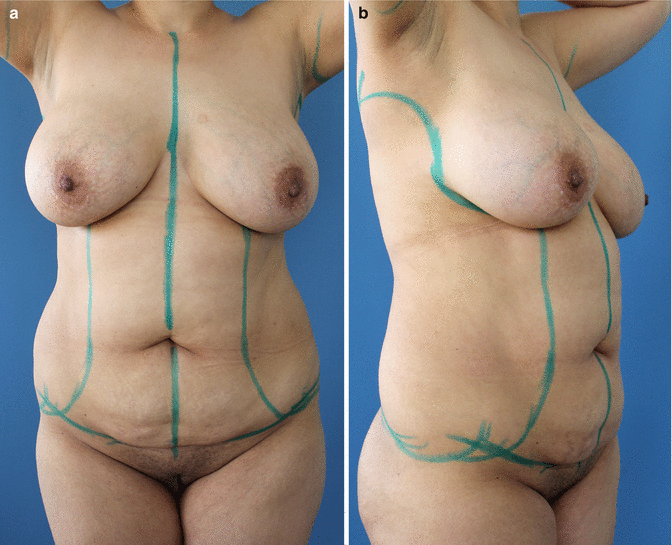

Fig. 11.2
(a, b) Abdomen is marked with all the lines necessary to perform the surgery. It is very important to mark the central abdominal midline, also the sidelines that would correspond to the outer edge of the rectus abdominal muscles. These sidelines are an extension of the mammary midline. Outside of these lines, we make a normal liposuction, while inside these lines, the abdominal flap is left a little bit thicker
In most patients, the anesthetic technique used is regional anesthesia with epidural block. This anesthesia gives us the advantage to avoid using lidocaine in the solutions that will be infiltrated into the subcutaneous tissue to achieve tumescent infiltration. This procedure allows the use of epidural catheter for postoperative analgesia during hospitalization. Intravenous (IV) fluids start before surgery, administering an average of one liter of 0.9 % normal saline with glucose 5 %. The surgery begins in the posterior region with the patient placed in prone position. The region is infiltrated with a solution consisting of a liter of saline 0.9 %, plus a milligram of epinephrine. The amount of infiltrated fluid is only the necessary to achieve tumescence of the subcutaneous tissue. Tumescence is only slightly higher than the super-wet technique, so that the range of infiltration to extraction is 1.2:1 approximately. With the patient in prone position, liposuction is performed in the back as needed, emphasizing a three-dimensional contour, particularly on waist and flanks. For liposuction, Mercedes-, cobra-, or Illouz-type cannulas are used. The lipoplasty is initiated in the deep plane with 4 mm diameter cannulas and finished with 3 mm cannulas. The fan technique is used for better homogeneity in the results and prevents irregularities; also we use the pinch test (Fig. 11.3) and observation of the thickness of the flap over the cannula to assess the regularity of the flap. At the end of surgery, drains are placed through intergluteal incisions; this is to facilitate liquid drainage (Fig. 11.4). Drains are removed about the 5th postoperative day. The main objective is to prevent seroma formation.
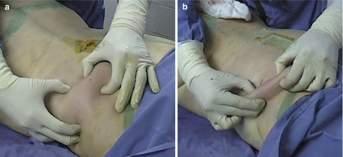
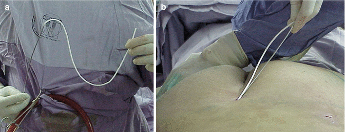

Fig. 11.3
(a) Pinch test prior to performing posterior liposuction. (b) Thinning flap in the posterior region after liposuction has been performed

Fig. 11.4
(a) Commonly soft silicone drains are used in areas where liposuction has been performed to facilitate drainage of liquid used in tumescence. (b) Drains are introduced using an infiltration cannula to reach up the top of liposuction
After the lipoplasty is finished in the posterior region, the patient is placed in supine position to begin surgery in the anterior region. In the anterior region, liposuction is performed outside of the lines that are defining the outer edges of the rectus abdominal muscles, using a technique similar to that described for the posterolateral region (Fig. 11.5). The pinch test (Fig. 11.3) is used and observation of the thickness of the flap over the cannula to assess the regularity of the flap and to determinate that the liposuction is completed (Fig. 11.6).
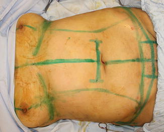
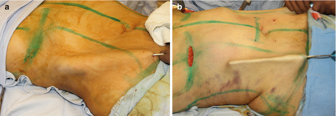

Fig. 11.5
Marking the abdominal area for abdominoplasty with the lines and delimitations

Fig. 11.6
(a) Lifting the cannula is also used to observe the thickness and regularity of the flap. Here is observed before liposuction. (b) After liposuction of the abdominal sides watching homogeneous and thinned flap
Subsequently, liposuction is performed on the central area of the abdomen. This central area is the area between the two outer lines corresponding to the rectus abdominal muscles. The technique used for central abdominal liposuction is done only in one direction, inferosuperior, in order to preserve at maximum the blood vessels. The approach is through a small incision in the abdominal flap to be resected during abdominoplasty (Fig. 11.7). The solution is infiltrated achieving tumescence in the manner previously described, to subsequently perform liposuction. The flap thickness of the central abdomen must be slightly thicker than the left in the lateral and posterior areas. Whereas in the central region, the flap thickness after liposuction must be between 3 and 4 cm in the lateral and posterior areas can be 2 cm thick. The type of incision used for abdominoplasty depends on the abdominal characteristics of each patient. A very important concept to note is that the skin retraction secondary to aggressive liposuction being performed in the abdominal region and lateral areas sometimes produces asymmetry in the position of the scar, and retraction may behave differently in each side. This is something that is difficult to control and is not dependent on the technique of the surgical procedure.
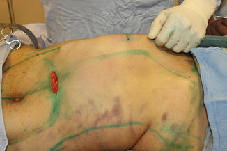

Fig. 11.7
Liposuction is also performed in the central abdominal flap that will be undermining during abdominoplasty. Liposuction is performed in one direction and a slightly thicker flap is left. This thick flap preserves vascularity and simulates the rectus abdominal muscles
Stay updated, free articles. Join our Telegram channel

Full access? Get Clinical Tree


