Chapter 41. Limited Incision, Nonendoscopic Foreheadplasty
David M. Knize, MD
INDICATIONS
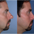
This limited incision foreheadplasty procedure is appropriate for any patient at any age that has ptotic eyebrows and skin line formation of the forehead and glabellar areas produced by underlying muscle hypertonicity. It is particularly indicated for the patient who has frontal scalp baldness yet has adequate temporal scalp hair to conceal incision scars.
PREOPERATIVE PREPARATION
With the patient looking into a mirror, elevate each eyebrow to the planned postoperative level and confirm that the eyebrow position and shape are consistent with the patient’s expectations. Evaluate the patient’s general medical status for undergoing surgery.
ANESTHESIA
This procedure can be done under conscious sedation or general anesthesia, either of which should be administered by an anesthesia provider.
POSITION AND MARKINGS
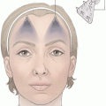
With the patient positioned supine, prep the face and scalp and apply a head drape. Mark the palpable superior temporal line of the skull (STL) on the forehead skin as a future reference point for several deeper structures (Fig. 41-1). Visualize the 6-mm wide zone of soft-tissue adhesion to frontal bone medial to the STL that requires complete release. Mark the approximate course of the deep division of the supraorbital nerve on the forehead skin. This nerve runs over periosteum between 0.5 and 1.5 cm medial to and parallel with the STL to supply sensation to the parietal scalp. Mark the suspension vector that will elevate the lateral eyebrow to the desired level, which is determined by elevating the lateral eyebrow using 1 finger. This vector line usually falls along the lateral side of the STL. At approximately 2 cm superior to the frontal hairline mark a 4-cm long line perpendicular the vector line, which will be the future temporal incision location on each side. The medial end of the incision should extend just over the STL; however, limit the extension to 0.5 cm medial to the STL to avoid transection of the deep division of the supraorbital nerve. No marking for medial eyebrow elevation is required, because elevation of the medial eyebrow is infrequently indicated. However, medial eyebrow elevation can be adequately obtained later, if necessary, by weakening the medial eyebrow depressor muscles. Finally, mark the area of excess eyelid skin in the usual manner for upper blepharoplasty, and then determine how much of this excess skin will be resuspended with the eyebrow elevation and mark that area for skin preservation.
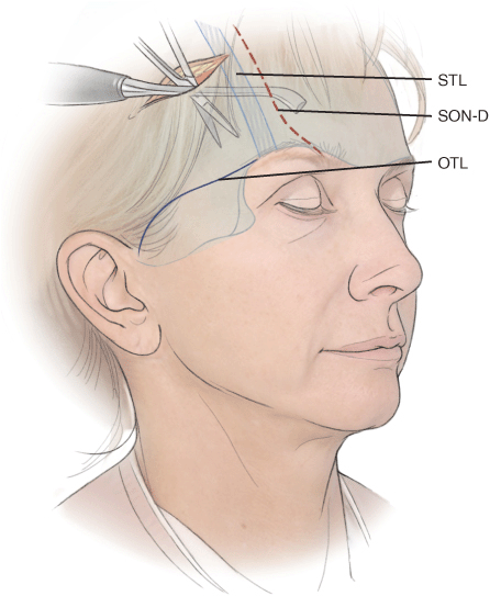
Figure 41-1 Soft-tissue release. The subperiosteal elevation extends across the frontal bone almost down to the level of the superior orbital rims. The temporal fossa dissection between the superficial and deep temporal fascial planes extends through the orbicularistemporal ligament (OTL) down to the zygomatic arch level. The superior temporal line of the skull (STL) and the usual course of the deep division of the supraorbital nerve (SON-D) are labeled. Note the zone of soft-tissue adhesion to bone medial to the STL (slant lines) that must be completely released to adequately transpose a forehead flap.
DETAILS OF THE PROCEDURE
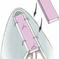
Infiltrate the forehead and temporal fossa areas with approximately 75 to 100 mL of 0.25% lidocaine with 1:400,000 epinephrine solution. The bilateral temporal scalp incisions are made as parallel to the axis of the hair shafts as possible to avoid injury to hair follicles. Initially, dissect between the superficial and deep temporal fascial planes (see Fig. 41-1
Related posts:


Stay updated, free articles. Join our Telegram channel

Full access? Get Clinical Tree


