Chapter 12 Lichenoid skin eruptions
Katta R: Lichen planus, Am Fam Physician 61:3319–3324, 2000.
Carrozzo M: How common is oral lichen planus? Evid Based Dent 9:112–113, 2008.
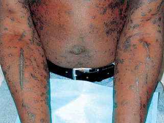
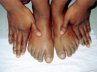
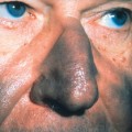
Primary lesions of the skin are 1 to 5 mm, flat-topped, violaceous, shiny papules (Fig. 12-1). While papules are often clustered, individual lesions tend to be discrete, with angulated (polygonal) borders. Wickham’s stria, a lacy white network present on the surface of the papules, is often of great diagnostic value.
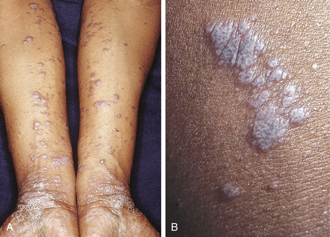
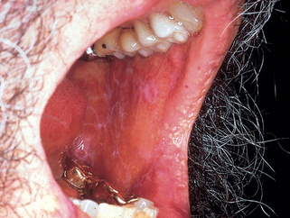
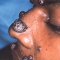
Figure 12-2. Lichen planus, showing reticulated leukoplakia of the buccal mucosa.
(Courtesy of James E. Fitzpatrick, MD.)
Dissemond J: Oral lichen planus: an overview, J Dermatol Treat 15:136–140, 2004.
Bruce AJ, Rogers RS 3rd: Lichenoid contact stomatitis, Arch Dermatol 140:1524–1525, 2004.