Chapter 46 Leukemic and lymphomatous infiltrates of the skin
Cutaneous Lymphoma Foundation. CTCL-MF fast facts. Available at: http://www.clfoundation.org/publications/publications.htm. Accessed December 6, 2006.
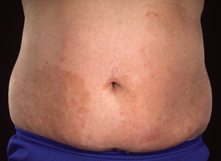
Large-plaque parapsoriasis presents as palm-sized or larger lesions located most frequently on the thighs, buttocks, hips, lower abdomen, and shoulder girdle areas (Fig. 46-2). The lesions may be pink, red-brown, or salmon-colored. They often have fine scale and show epidermal atrophy with cigarette-paper wrinkling. Some patients may have lesions with a netlike or reticular pattern with telangiectasia and fine scale. This clinical type of lesion is referred to as retiform parapsoriasis or poikiloderma atrophicans vasculare. Between 15% and 20% of patients with large-plaque parapsoriasis eventually develop mycosis fungoides.

Figure 46-2. Large-plaque parapsoriasis. The lesion was unresponsive to topical treatment.
(Courtesy of the Fitzsimons Army Medical teaching files.)
Sehgal VN, Srivastava G, Aggarwal AK: Parapsoriasis: a complex issue, Skinmed 6:280–286, 2007.
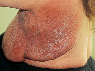
A rare variant of mycosis fungoides is granulomatous slack skin disease (Fig. 46-3). This disorder is characterized by the slow development of lax erythematous skin that eventually develops large pendulous folds of redundant integument. Histologic examination shows a dense atypical granulomatous infiltrate with destruction and phagocytosis of elastic tissue.


