Ablative laser resurfacing has been the gold standard throughout the 1980s and 1990s for improving skin texture by renovating photo damaged and aging skin.
Although excellent results have been obtained with the use of ablative laser devices, the use of non-ablative laser systems have been increasingly popular as an alternative to obtaining the same results.
The mechanism of ablative laser resurfacing involves the removal of the epidermis and the dermis, causing a wound to develop which may take up to several months to heal.
The use of non-ablative laser systems have minimal side-effects by causing direct thermal damage to the dermis, while sparing the epidermis, and reducing the risks of post-treatment adverse effects and downtime.
Indications and Contraindications – Clinical information regarding these devices with published evidence of safety and efficacy
Introduction
The field of sub-surfacing lasers is also known commonly by the terms non-ablative laser resurfacing, photorejuvenation, and laser toning. A variety of lasers and light sources have been developed and utilized for sub-surfacing and will be the subject of this review.
Included in the overall category of sub-surfacing lasers are a diverse range of technologies which include the KTP lasers at 532 nm; the pulsed dye lasers at 585–595 nm; the Nd:YAG lasers which encompass the 1,064 nm long pulsed systems, the 1,064 nm Q-switched laser systems, and the 1,319/1,320 nm laser systems; the 1,450 nm diode laser systems; the erbium glass laser systems at 1,540 nm; as well as the spectrum of intense pulsed light (IPL) systems at a wavelength range of 500–1,200 nm. All of these medical devices have proven successful for non-ablative laser/light source resurfacing, photorejuvenation, and the improvement of acne scars.1,2
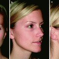
These devices were developed, from a historical point of view, to rival the ablative laser resurfacing systems which enjoyed much popularity in the 1980s and 1990s. The ablative laser systems, predominantly identified by the erbium laser systems at 2,940 nm and the carbon dioxide resurfacing lasers with a wavelength of 10,600 nm, were and are the mainstays of the ablative laser resurfacing systems. The ablative laser resurfacing systems are the gold standard for skin resurfacing and still to this day are the standards to which all other laser or light sources for rejuvenation of the skin are judged as to their safety and efficacy. The ablative laser resurfacing systems work by removing the entire epidermis and portions of the dermis which results in a wound that, upon healing, gives a desired rejuvenation effect. These devices have been used for many years and have proven effective in improving skin roughness, fine lines and wrinkles, and skin dyschromias which are common manifestations associated with skin aging and photodamage. These ablative laser resurfacing devices can produce very nice and long-lasting results, as evidenced in Figs. 1 and 2. However, when performing these types of laser procedures, patients must understand that there will be significant downtime with these treatments, lasting upwards of 1 week or more before full reepithelization will occur, and then several more weeks where the skin is still pink until complete healing has occurred. As well, some patients have prolonged erythema, as seen in Fig. 3, which requires appropriate and thoughtful intervention from qualified clinicians to help minimize. And, as a long term sequelae to many of these procedures, a significant number (reported anywhere from 10–20%) may develop a post-treatment hypopigmentation, which may not be evident for several months to years following the initial procedure, as depicted in Fig. 4. However, these ablative laser resurfacing devices still remain the gold standard for skin resurfacing but other devices were developed in an attempt to reproduce these kinds of results, with minimal downtime, predictable results, and less chances for adverse or long term results.3–5
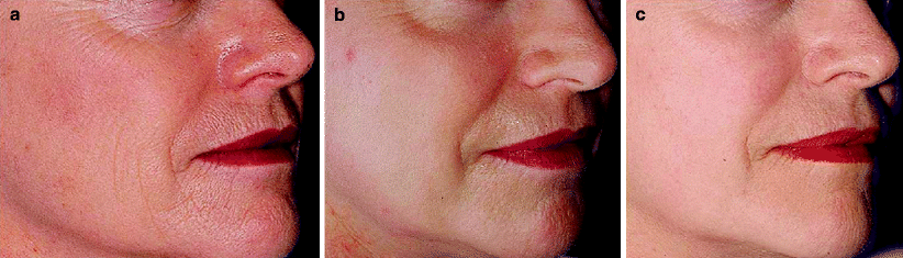
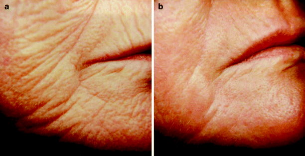
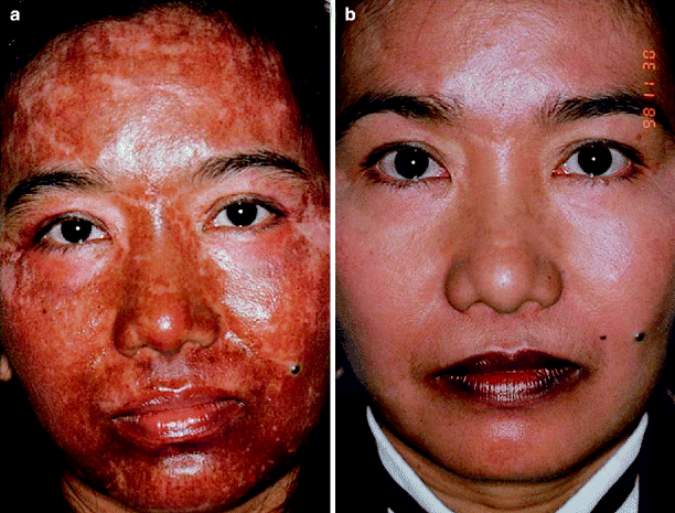
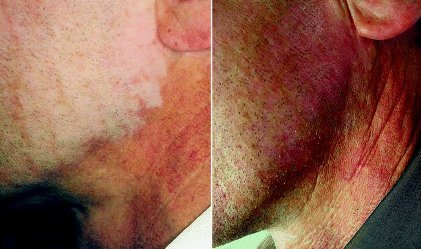
Fig. 1
(a–c) Clinical examples of ablative laser resurfacing with long-lasting results
Fig. 2
(a, b) Clinical examples of ablative laser resurfacing with long-lasting results
Fig. 3
(a, b) Clinical example of prolonged erythema after ablative laser resurfacing
Fig. 4
Clinical example of long-term sequelae after ablative laser resurfacing
The purpose of the development of the non-ablative laser resurfacing is to affect changes in the skin similar to what the ablative laser resurfacing devices achieve. These devices were developed to improve the skin’s texture, to improve facial lines and wrinkles and surface irregularities including facial scars. In addition, some of these devices also address pigmentary dyschromias and vascular changes in the skin associated with photodamage and actinically damaged skin. The non-ablative lasers and light sources work via thermal injury to the dermis, with epidermal sparing, thereby reducing the potential for any associated with these procedures. These medical devices have been demonstrated to work over a period of time and through a series of treatments, usually in the realm of 5–6 treatments given at 3–4 week intervals.3 Maintenance therapy, although not addressed in many accounts of sub-surfacing lasers, is usually required at certain time intervals, as well as proper skin care to maintain the rejuvenation effect one is attempting to achieve. In contrast to the amount of literature which the ablative laser resurfacing devices have enjoyed in the medical literature, these sub-surfacing lasers have not had an overwhelming scientific base in the literature but have had descriptive coverage of the various devices and their effects.
Various classifications of these sub-surfacing lasers have been used over the past several years – we will classify them as shown in Table 1. Table 1 shows that these medical devices can be classified into (1) vascular lasers, (2) mid-infrared lasers and (3) intense pulsed light sources. The remainder of this chapter will review the literature of the vascular lasers, the mid-infrared lasers, and the IPLs.
Table 1
Sub-surfacing lasers
Device type | Device name | Manufacturer |
|---|---|---|
(1) Vascular lasers | ||
KTP lasers | Aura (laserscope) | Iridex http://www.iridex.com/ |
Viridis derma | Quantel medical http://www.quantelmedical.com/ | |
Pulsed dye lasers | V-beam/ | Candela http://www.candelalaser.com/ |
Veam perfecta | Candela http://www.candelalaser.com/ | |
V-star | Cynosure http://www.cynosure.com/ | |
Cynergy | Cynosure http://www.cynosure.com/ | |
NLite | ICN Pharmaceuticals http://www.yestheyrefake.net/NLite.htm | |
(2) Mid-infrared lasers | ||
Q-switched 1,064 nm | Medlite C6/ | Hoya ConBio http://www.conbio.com/ |
RevLite | ||
Long-pulsed 1,064 nm | YAG 5 | Palomar http://www.palomarmedical.com/ |
Gentle YAG | Candela http://www.candelalaser.com/ | |
Apogee Elite | Cynosure http://www.cynosure.com/ | |
1,319/1,320 nm | Profile 1,064 nm | Sciton http://www.sciton.com/ |
CoolTouch® C3 | CoolTouch® Corp http://www.cooltouch.com/ | |
1,450 nm | Profile 1319 | Sciton http://www.sciton.com/ |
1,540 nm | SmoothBeam | Candela http://www.candelalaser.com/ |
Aramis | Quantel http://www.quantelmedical.com/ | |
(3) Intense pulsed light sources | Lumenis one™ | Lumenis http://www.lumenis.com/ |
BBL | Sciton http://www.sciton.com/ | |
PhotoSilk | Cynosure http://www.cynosure.com/ | |
Harmony | Alma lasers http://www.almalasers.com/ | |
PPX/isolaz | Aesthera http://www.aesthera.com/ | |
Ellipse flex | ||
Studies have shown that KTP lasers have better collagen formation results when compared to 1,064 nm lasers in the treatment of skin photo-rejuvenation. Pulsed dye lasers (PDL) function best in the treatment of vascular lesions (i.e., port wine stains and hemangiomas) with significant production of procollagen type I and type III. Increased activity of dermal fibroblasts and mucin, as well as the thickening of the stratum spinosum in the dermis, has been noticed in the restoration of degenerated skin. The use of modern PDL systems for skin rejuvenation provides non-ablative results by minimizing side-effects and reducing purpura. |
Vascular Lasers
KTP Lasers
The KTP lasers have traditionally been utilized to treat small caliber facial telangiectasias on the face and there is little in the medical literature which can be attributed to the use of the KTP lasers in rejuvenation of the skin. In a study by Lee,6 150 patients were treated with the KTP laser and the 1,064 nm laser separately and together for non-ablative facial rejuvenation. Patients in this series received 3–6 treatments of both the KTP laser alone, the 1,064 nm laser alone, and the combination of both devices. Skin biopsies looking at histologic effects in the skin were performed at 1, 2, 3, and 6 months following the last treatment. The results showed that the KTP laser was superior to the 1,064 nm laser in improving the parameters of photorejuvenation of the skin but that the combination of devices was superior to either one of the devices used alone. Post-therapy skin biopsies confirmed the presence of new collagen formation as well as a result of these lasers systems. Others, including Goldberg7 found similar results in ten patients – again treated with a millisecond Nd: YAG laser and a millisecond KTP laser showed greater effects on tissue rejuvenation. Tan et al.8 also demonstrated this synergistic effect of these two laser systems – 25% more improvement with the combined systems at 1 month for more than one third of the patients which increased to 40% at the end of 4 months.
Pulsed Dye Lasers
Pulsed dye lasers are also known commonly as flashlamp-pumped pulsed dye lasers are the prototype medical devices that were developed which adhere to the principle of selective photothermolysis.9 Selective photothermolysis is a principle which stated that a specific wavelength of light can specifically destroy a chromophore within the skin, which in the case of the pulsed dye lasers is hemoglobin. The pulsed dye laser systems have improved in many facets over the years, especially in their methods of epidermal cooling and in that they have longer pulse durations than their predecessor devices, which reduces the incidences for purpura as a result of the therapy, considered one of the necessities of the early pulsed dye lasers but also one of the drawbacks from the treatment. These devices have been routinely used to treat vascular lesions (facial telangectasia, diffuse erythema, and other superficial vascular lesions) and their successes in the treatment of port wine stains and hemangiomas have been reviewed in the medical literature.10–12 Pulsed dye lasers have shown to be useful in reducing the erythema and improving stretch marks13 and these devices have also been used successfully to treat hypertrophic scars and keloidal lesions, especially being able to reduce the associated erythema sometimes associated with these lesions.14,15 In the process, many of these scars have shown dermal remodeling and an associated shrinkage of the scar itself. The exact mechanism of pulsed dye induced collagen formation is not clear but it has been proposed that thermal induced damage to vascular endothelium may produce cytokines leading to dermal remodeling and thus the improvement of fine lines and wrinkles.3
Several clinical evaluations have been performed which support the use of the pulsed dye lasers for a non-ablative rejuvenation effect. Rostan et al.16 utilized a long pulsed 595 nm laser in evaluating wrinkles of the cheeks. Fifteen patients received a series of four treatments at monthly intervals, either with the laser on or with cryogen only in the control group. The group performed skin biopsies prior to the pulsed dye laser treatments and at 4–6 weeks following the last laser treatment, as well as 3 months following the last laser treatment. They specifically looked at routine histology, procollagen I production and the activity of dermal fibroblasts. They found that the Grenz zone was moderately thicker in 50% of patients in both groups treated, but the degree of thickening was greater in the treatment groups than the controls. Similar findings were also seen at 3 months following the laser treatments, when an increase in dermal collagen was also observed in the treatment sites. A statistically significant improvement in the clinical grading scale for photodamage was also noted in the study.
Zelickson et al.12 evaluated ten patients with mild to moderate facial lines and wrinkles and ten patients with moderate to severe facial lines and wrinkles that were treated with a single laser treatment with a 585 nm pulsed dye laser with a 450 um pulse duration. Clinically 9 out of 10 patients in the mild to moderate wrinkle group showed 50% or more improvement with 3/10 showing 75% or more improvement. All of the patients maintained their improvements for 6 months following their last laser treatment. In contrast, only 3/10 patients in the moderate to severe wrinkle group improved during the study. Skin biopsies from those patients in the mild to moderate group were performed at 6 and 12 weeks following the laser treatment. The biopsy specimens showed a thickened stratum spinosum and a thickened collagen layer in the superficial dermis as well as increased mucin in the superficial dermis. They also performed ultrastructural evaluations which demonstrated an increase in collagen fibers and an increase in the number of fibroblasts in the treated skin. They also noted that there was an increase in normal appearing elastic fibers and a decrease in the clumping of degenerated elastic fibers in the skin. Zelickson and Kist17,18 have also reported results from skin biopsies performed after two periorbital 585 nm pulsed dye treatments or IPL treatments (6 week apart) in which in-situ hybridization mRNA probe studies were performed 6 weeks after the second treatment. These studies showed an 18% increase in type I collagen production after the IPL treatments compared to a 23% increase in type I collagen production after the pulsed dye therapy. Collagenase transcriptase activity was 32% for the IPL and 40% for the pulsed dye laser. The authors concluded that the observed increases in mRNA in fibroblasts indicated that through this non-ablative light, extracellular matrix protein production by dermal fibroblasts is increased, thus resulting in the effects seen.
Others have also looked at the pulsed dye lasers for its potential in rejuvenation of the skin. Bjerring et al.19 utilized a 585 nm pulsed dye laser with a 350 um pulse duration and studied suction blisters which had been treated with the pulse dye laser. Using special markers for the production of type III procollagen they were able to demonstrate significant increases in type III procollagen levels after a single treatment with the pulsed dye laser, but also demonstrated a drop in type III procollagen levels after a retreatment. Goldberg et al.20 also performed skin biopsies with a similar pulsed dye laser looking at facial lines and wrinkles. In this study, patients received two pulsed dye laser treatments. Pre-treatment and 6 month post-treatment clinical evaluations and skin biopsies were analyzed. Forty percent of the individuals noted mild improvement in their wrinkle appearance. Ultrastructural analyses showed increases in type I and III collagen in those treated with the pulsed dye lasers. A second study by these authors21 evaluated clinical, histologic, and ultrastructural changes after non-ablative treatments utilizing varying settings in the same subject. Ten patients were included in this clinical trial. The patients were randomly divided into two groups of five. All of the patients received 595 nm pulsed dye laser treatments to the periorbital areas. The first group was treated once, while the second group was treated twice at a 1 month interval. Each side of the face was treated with distinctly different laser settings. Seventy percent of the patients noted mild to moderate improvement clinically after treatment at 6 months following the last treatment. There was no difference between receiving one or two treatments in this study. Histologic and electron microscopy improvement was noted in all of the patients.
Examples of improvement in rejuvenation as a result of pulsed dye lasers are shown in Figs. 5 and 6.
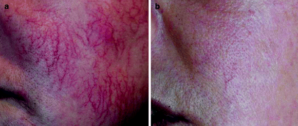
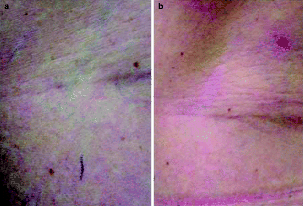
Fig. 5
(a, b) Clinical example of rejuvenation following pulsed dye laser treatment; 10 msec, 7 J/cm2, Delay short, Nd:YAG 10 ms, 40 J/cm2 (Photos courtesy of Cynosure/P. Boixeda M.D.)
Fig. 6
(a, b) Clinical example of rejuvenation following pulsed dye laser treatment; Post 2 treatments, PDL 40 ms, 11 J/cm2, Delay Medium, Nd:YAG 15 msec, 60 J/cm2 (Blue reticular vein.) (Photos courtesy of Cynosure/R. Adrian M.D.)
Related posts:


Stay updated, free articles. Join our Telegram channel

Full access? Get Clinical Tree


