Laser treatment of ethnic skin

10 Laser treatment of ethnic skin


Introduction


The evolution of population demographics in the 21st century is such that patients with ethnic skin will become of increasing importance to any practicing dermatologist. Ethnic skin comprises a diverse group, including Chinese, Japanese, Indian, Pakistani, Hispanic, Latino, African, Afro-Caribbean, and African American. Their skin color typically falls within the Fitzpatrick’s skin phototypes III–VI.


Ethnic skin differs from the Caucasian counterpart in several aspects. The larger melanocytes in skin of color produce more melanin and melanosomes are distributed individually in keratinocytes, conferring significant photoprotection. Photoaging tends to manifest more frequently as pigmentary changes, as opposed to wrinkle formation. Congenital and acquired pigmentary disorders such as nevus of Ota, Hori’s macules, and melasma are also more commonly encountered. Management of pigmentary conditions is therefore an important concern for most patients with ethnic skin. However, the high melanin content in such skin, coupled with the broad absorption of melanin on the electromagnetic spectrum, can often present therapeutic challenges during laser treatment, as a competing chromophore to the intended pigmented target is present throughout ethnic skin. Post-inflammatory hyperpigmentation (PIH), rarely seen in light-colored skin, can be a common outcome during inexperienced laser use in darker skin types. This chapter aims to discuss the effective and safe use of laser and light sources in the management of common conditions seen in ethnic skin, in order to optimize results and minimize complications. We will also highlight good practices to adopt, and pitfalls to avoid.


Evaluating the patient with ethnic skin


A thorough history and examination are essential in establishing a correct diagnosis prior to treatment. Standardized digital photography is helpful in recording the baseline appearance, and any subsequent improvement. Additional aids such as UV photography or Wood’s light can be useful, for example, in assessing the epidermal and dermal components of melasma. The patient’s treatment objective and expectation should be enquired, and treatment options and associated risks discussed. Providing additional printed material regarding suggested procedures is often helpful. Obtaining informed consent prior to procedure is essential and good practice. Establishing realistic patient expectations through good rapport is imperative in achieving a satisfactory outcome.


A detailed patient interview can identify the patient’s concern and highlight the primary problem, which must be addressed. For example, a patient presenting for management of acne PIH and scarring will require treatment for their active acne before embarking on treatment of the secondary complications. Any relative contraindications to laser and light treatment should be excluded, such as infection or recent sun exposure of the treatment site, isotretinoin use in the last 6 months, photosensitivity or use of photosensitizing drugs, immunocompromised state, predisposition towards keloid or hypertrophic scarring, pregnancy, and personal or family history of melanoma. The patient’s Fitzpatrick’s skin type should be noted.


Taking a thorough medical history also helps clinicians identify patients who have unrealistic expectations, or psychiatric disorders such as body dysmorphic disorders (BDD). Such patients have an excessive preoccupation with an imagined or slight defect in appearance, which results in a clinically significant distress and impairment in functioning. A recent study reviewing 401 adults with BDD symptoms in an ethnically diverse group found significant differences between Asians and Caucasians, with Asians reporting more concerns with straight hair and dark skin, and less concerns about body shape. A simple and reliable questionnaire for the diagnosis of BDD has been developed by Dufresne and colleagues, and can be a useful adjunct in suspected cases. BDD is a contraindication to laser treatments, and these patients should be referred for psychiatric and psychotherapeutic treatments.


Obtaining written consent with a clear outline of risks and benefits is essential and protects both the clinician and the patient. The clinician should ensure that patients have a good understanding of different treatment options, the expected outcome, the associated downtime, postoperative care, and potential risks from the procedure. There should be adequate opportunity for the patient to have all their questions answered.


Any sun exposure during several weeks prior to laser treatment can predispose patients with ethnic skin to a higher risk of post-inflammatory hyper or hypopigmentation. The use of sunscreens and topical bleaching agents for at least 2 weeks prior to any such treatment can help reduce the risk of PIH. Patients should be provided written guidance regarding postoperative care, with additional emphasis on sun protection and avoidance for at least 4 weeks after.


Treatment of epidermal pigmentation


Freckles and lentigines


Freckles and lentigines are benign pigmented lesions commonly seen in ethnic skin. Freckles occur in adolescence and are relatively uniform in distribution, size and color. Histologically, they are characterized by epidermal hypermelanosis without an increase in melanocyte numbers. Lentigines tend to appear later and vary in size, color and distribution. Histologically, both epidermal hypermelanosis and increase in melanocyte numbers are seen. Epidermal rete ridges are also elongated and clubbed.


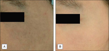
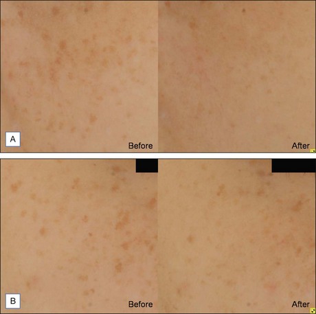
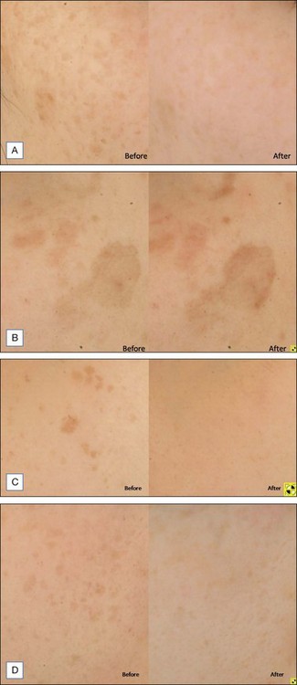
As melanin has a broad absorption spectrum ranging from 250 to 1200 nm, various lasers have been used to target cutaneous pigmentation, usually with excellent results. Anderson et al were the first to demonstrate the effectiveness of Q-switched (QS) lasers in the treatment of cutaneous pigmentation. Frequency-doubled QS neodymium : yttrium-aluminum-garnet (Nd : YAG), QS ruby, and QS alexandrite lasers with respective wavelengths of 532 nm, 694 nm, and 755 nm, have all been used with good results in lighter-skinned patients but PIH risk of 10–25% has been reported when used on ethnic skin. Chan et al compared 532 nm frequency-doubled Nd : YAG lasers with different pulse durations in the treatment of facial lentigines in Chinese patients, and found similar efficacy between QS 532 nm Nd : YAG and long pulsed (LP) 532 nm Nd : YAG, but a higher risk of postoperative hyperpigmentation with the QS device. A recent study by our group comparing QS and LP alexandrite for the treatment of freckles and lentigines in 20 Chinese patients showed similar results (Fig. 10.1). There was significant improvement in pigmentation in both groups, with no difference between the groups. However, the risk of PIH was 22% in the QS group, compared with 6% in the LP group. Patients also complained of more severe pain, erythema, and edema with the QS device. These findings were further validated in a retrospective study due to be published, comparing treatment of lentigines in 40 Chinese patients with four different devices; 595 nm long pulsed dye laser (LPDL), 755 nm alexandrite laser, 532 nm QS Nd : YAG, and 532 nm LP potassium-titanyl-phosphate (KTP) laser (Fig. 10.2). The results showed that a long pulse laser and small spot size appear to reduce the risk of PIH in darker skin types (see Case study 1 at end of chapter).


LP lasers, with their longer millisecond pulse width, result in more absorption by target melanin and less absorption by competing chromophores such as oxyhemoglobin, and surrounding pigment-laden skin. This is particularly important in reducing the risk of PIH in ethnic skin. The postulated reason is that LP lasers cause melanin destruction by photothermolysis only. In contrast, QS lasers emit high-energy, nanosecond radiation, causing both photothermal and photomechanical effects. Not only is the target pigmented lesion destroyed by the short burst of intense radiation, but surrounding melanin and oxyhemoglobin are also damaged, resulting in altered activity of melanocytes, hemosiderin deposition from damaged vessels, and subsequent PIH.


Spot sizes are also an important consideration when treating darker skin types. From our retrospective study comparing four different laser devices above, we noted that when using LP alexandrite in the treatment of epidermal pigmentation in Asian skin, despite its long pulse width, significant improvement in lesional pigmentation was not found, and was associated with the highest risk of PIH (20%). We postulated that the large spot size (10 mm) in the LP alexandrite laser may have led to inadvertent treatment of surrounding unaffected skin, when the lesion treated is smaller than the spot size available, and this is especially pertinent in ethnic skin where contrast between the lesional and non-lesional skin is low.


There is increasing evidence to support the use of diascopy during laser treatment to reduce the risk of PIH. This is especially true when using pigment lasers that target both oxyhemoglobin and melanin such as the LPDL. Compression of the skin surface by the flat glass window on the handpiece leads to emptying of blood vessels, reducing the risk of vascular damage, and subsequent purpura, hemosiderin deposition, and PIH. The effectiveness of such simple diascopy is supported by results from studies carried out by Kono et al, who conducted different studies using 595 nm LPDL, repeatedly demonstrating its effectiveness and safety when used with compression for the treatment of lentigines in ethnic skin, when compared with IPL and QS ruby laser. Our retrospective study also showed that 595 nm LPDL and 532 nm LP KTP, both of which utilize a compression window, have better results and less complications compared with 532 QS Nd : YAG and 755 nm LP alexandrite laser.


Intense pulsed light (IPL) sources emit a broadband of visible light (400–1200 nm) from a non-coherent filtered flashlamp and target melanin through photothermal effects. Negishi et al carried out two studies looking at photorejuvenation using IPL. Results from the first study involving 97 Asian patients showed that more than 90% reported a reduction in pigmentation after three to six treatments at intervals of 2–3 weeks (cutoff filter 550 nm, 28–32 J/cm2, double pulse mode of 2.5–4.0/4.0–5.0 ms, delay time 20/40 ms). The second study used IPL with an integrated cooling system and found 80% of the 73 patients had a significant reduction in pigmentation after three to five treatments at intervals of 3–4 weeks (cutoff filter 560 nm, 23–27 J/cm2, double pulse mode of 2.8–3.2/6.0 ms, delay time 20/40 ms). Kawada et al also treated lentigines and freckles in 60 patients with IPL and reported more than 50% improvement in 68% of these patients after three to five treatments at intervals of 2–3 weeks. No PIH was reported in any of these studies. A further split face study comparing QS alexandrite and IPL for freckles and lentigines in Asians found significant improvement with the QS device. However, the risk of PIH was also higher with the QS device, especially in patients with lentigines. No PIH was seen in the IPL group. These studies serve to highlight IPL, through photothermal effects, is effective and safe in the treatment of epidermal pigmentation in ethnic skin. However, multiple treatments are required. Furthermore, with a large spot size, if the contrast between the lesional and non-lesional skin is low, the therapeutic window is much narrower. Either the operator uses suboptimal energy leading to minimal effectiveness, or above threshold energy that may lead to greater risk of injury to surrounding normal skin.


A treatment algorithm for epidermal pigmentation is as follows:


Treatment of dermal pigmentation
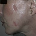

Dermal pigmentation such as Nevus of Ota and acquired bilateral nevus of Ota-like macules (ABNOM) or Hori’s macules are much more commonly encountered in ethnic skin. Removal of unwanted tattoos and hair require the removal of dermal pigment. In such conditions, devices with longer wavelengths, and hence deeper penetration, are utilized to provide enhanced clearance.


Nevus of Ota


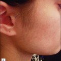
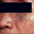
Nevus of Ota is an oculodermal melanocytosis affecting approximately 0.6% of the Asian population at birth or developing in their teens. Clinically, it presents as a bluish-black hyperpigmentation along the ophthalmic or maxillary branches of the trigeminal nerve. QS lasers, including QS ruby, QS alexandrite, and QS 1064 nm Nd : YAG, have all been used to achieve good therapeutic results.Watanabe and Takahashi looked at 114 nevus of Ota patients treated with QS Ruby and reported a good to excellent degree of lightening after three or more sessions. Kono et al confirmed the findings when he reviewed 101 nevus of Ota patients treated with QS Ruby and reported 56% achieving 75% improvement, and 36% achieving complete clearing. Hypopigmentation was seen in 17% of patients and hyperpigmentation in 6%. Studies comparing the use of QS alexandrite and QS 1064 nm Nd : YAG found the former better tolerated but the latter more effective after three or more treatment sessions. The longer wavelength of the QS Nd : YAG targets dermal pigment effectively with minimal epidermal damage in darker skinned individuals (Fig. 10.3). More recently, fractionated laser technology has been added to the currently available options. Near-complete clearance of a case of nevus of Ota after serial therapy using 1064 nm QS Nd : YAG, followed by 1550 nm non-ablative fractionated erbium-doped fiber laser treatments at 2-month intervals; and a further case achieving complete clearance with sequential same day therapy with the same devices have been reported. Treatment of nevus of Ota at a younger age requires fewer treatment sessions and is associated with fewer complications compared with older patients, and early treatment is therefore recommended (see Case study 2). It is important to note that the risk of recurrence is estimated to be between 0.6% and 1.2%8, which has important implications particularly when counseling pediatric patients.


Apr 2, 2016 | Posted by in Laser surgery | Comments Off on Laser treatment of ethnic skin

Full access? Get Clinical Tree

Get Clinical Tree app for offline access