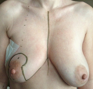
Fig. 29.1
Preoperative image showing palpable mass (circled) and the microcalcification (marked with clip and wire)
29.2 Surgery
Figure 29.2 shows preoperative pictures after guided wire localization and preoperative drawings for an inverted-T reduction mammoplasty technique. Figure 29.3 shows the breast completely freed from the skin and the two defects after bisegmentectomy laterocranial and laterocaudal in the right breast. The sutures are used to close the defect in the upper outer quadrant of the breast. Figure 29.4a also shows both defects without defect correction, and Fig. 29.4b demonstrates the breast reshaped after parenchymal sutures.


