2
Intense Pulsed Light for Full Facial Rejuvenation
Nonablative broadband light and laser devices have grown in popularity in recent years. One such device, the intense pulsed light (IPL), was introduced in the mid-1990s and has become a well-recognized treatment modality to improve the appearance of the photoaged face. Photoaging consists of the characteristic clinical changes to facial and other sun-exposed skin caused by chronic exposure to ultraviolet (UV) light. These changes include the appearance of telangectasias, rough texture, fine and coarse wrinkles, yellow or sallow color, and mottled pigmentation.1
The overall appearance of aging skin is primarily related to the quantitative effects of sun exposure over time. UV radiation damages structural components of the dermis, chiefly collagen and elastic fibers. A common pathway whereby photoaging occurs is through UV-generated reactive oxygen species.2 Facial appearance, however, is also affected by genetic factors, intrinsic factors, disease processes such as rosacea, and the overall loss of cutaneous elasticity associated with age. Signs of photoaging are becoming more evident in increasingly younger individuals, especially those regularly engaged in outdoor professions or recreational activities, as a consequence of exposure to increasingly high levels of UV radiation.3 Treatment of these changes with nonablative devices has been termed photorejuvenation and can be approached in a rational, stepwise fashion (Table 2-1).
Nonablative laser devices classically emit energy in coherent wavelengths within the near infrared to infrared spectrum (Table 2-2). Intradermal water is the target chromophore and heat generated by this interaction causes thermal injury within the dermis, initiating a cascade of molecular repair events that promote collagen formation and deposition.4 Clinically, this is manifested by subtle improvements in fine wrinkling and skin texture.
Alternatively, vascular-specific laser devices with wavelengths from 585 to 600 nm are also thought to cause thermal injury to dermal elements. These lasers are commonly used to treat the telangiectatic component. Energy is selectively absorbed by the target chromophore, hemoglobin. Light energy is converted to heat, which damages the vessel walls and radiates into the surrounding dermis, causing injury and repair with the formation of new collagen.5
By contrast, IPL is one proprietary noncoherent device in a class of several such devices, which emits a spectrum of wavelengths in the 515 to 1200 nm range. It allows for variation of pulse length/mode, delay between pulses, and fluence energy. Cutoff filters eliminate the lower range of wavelength of light emitted. Consequently, patients of darker skin types (Fitzpatrick III–IV) can be safely treated. Target lesions include blood vessels as well as lentigines. Selective thermal heating of chromophores including hemoglobin, water-containing tissue (collagen), and melanin is thought to induce neocollagenesis.6 Intrinsic cooling provided by IPL devices allows for repair of photoaging structural changes in the skin without disruption of cutaneous integrity. The resulting patient treatment has a low-risk profile and minimal downtime.
 Technology
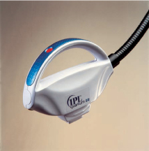
IPL devices consist of a flashlamp housed in an optical treatment head, in which reflecting mirrors are water cooled (Fig. 2-1). Optically coated quartz filters are placed over the window of the optical treatment head to eliminate wavelengths lower than the filter. For faster recycling times, certain optical heads have water circulating around the flashlamp. The filter crystals must be optically coupled to the skin with a water-based gel.3
| Lesion | Device |
| Facial telangiectasias | IPL, PDL |
| Mottled pigmentation | IPL, PDL, Microdermabrasion |
| Mild rhytids | IPL, CT2 |
| Moderate rhytids | CT2 |
IPL, intense pulsed light; PDL, Pulsed dye laser (595 nm); CT2, CoolTouch II Laser (1320 nm)(Cool Touch Corp., Roseville, California).
When filtered, the IPL device is capable of emitting a broad bandwidth of light from 515 to ∼1200 nm. This bandwidth is modified by the application of filters, which exclude wavelengths lower than the filter to help protect against epidermal heating. Although the output is not uniform across this spectrum, it has been shown that during a 10 msec pulse relatively high doses of yellow light at 600 nm are emitted, with far less red and infrared, although output clearly is demonstrated beyond 1000 nm.3
Filters applied for vascular lesions are 515, 550, 570, and 590 nm. Longer filters 615, 645, 695, and 755 nm, which cut off much more of the yellow wavelengths, are used for various applications but most commonly for photoepilation. Importantly, IPL involves not only filtering and eliminating the lowest wavelengths emitted by the flashlamp but also the ability to easily manipulate pulse durations and to couple these pulse durations with precise resting or thermal relaxation times.

Figure 2-1
Stay updated, free articles. Join our Telegram channel

Full access? Get Clinical Tree


