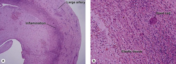
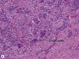
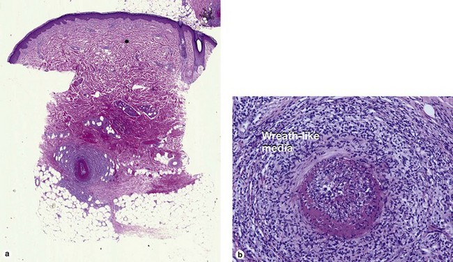
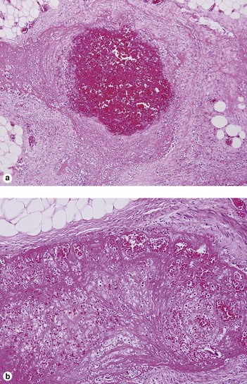
Inflammatory vascular diseases

Chapter 11


Inflammatory vascular diseases


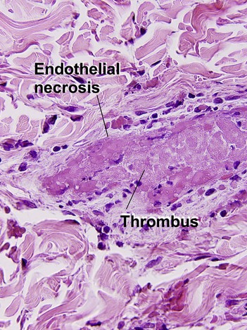
Leukocytoclastic vasculitis (LCV)


Clinical lesions of leukocytoclastic vasculitis are purpuric, and often palpable. Vasculitis involving arterioles commonly produces livedo reticularis or stellate infarcts.


Large vessel vasculitis


When a large vessel is involved by vasculitis, it is critical to determine whether the involved vessel is an artery or a vein. Arteries are characteristically round, with a wreath-like muscularis and an internal elastic membrane. Veins are characteristically oval, with a bundled muscularis. They may have visible valves, and lack an internal elastic membrane. So-called “arterialization” of veins occurs when they are subjected to elevated hydrostatic pressure. This phenomenon is occasionally noted in cutaneous vessels, but is best demonstrated in coronary artery bypass grafts. The grafted vein develops a prominent internal elastic membrane, but retains the bundled muscularis characteristic of a vein.


Giant cell arteritis (temporal arteritis)


Temporal arteritis often involves the vessel in a focal, beaded fashion, so an adequate length of temporal artery (ideally 2 cm) should be submitted for examination.


Polyarteritis nodosa


Polyarteritis nodosa commonly presents with livedo reticularis and subcutaneous erythematous or hyperpigmented nodules. The biopsy typically demonstrates neutrophilic vasculitis involving an artery within the subcutaneous fat. Surrounding lobular necrosis is present.


Medium vessel vasculitis


These diseases are characterized by leukocytoclastic vasculitis involving vessels larger than the post-capillary venule. The endothelium is frequently necrotic. The biopsy demonstrates a superficial and deep perivascular infiltrate with neutrophils, karyorrhexis, expansion of the vessel wall, fibrin deposition within the vessel wall, and erythrocyte extravasation.


Wegener’s granulomatosis


Wegener’s granulomatosis commonly involves the upper airway. The skin of the nose may become necrotic. Skin lesions may occur in other locations, especially the extremities. The histologic pattern is that of a “big 5” vasculitic disorder. Individual vasculitic foci may evolve into stellate abscesses (palisaded granulomas with a central stellate collection of neutrophils). Multinucleate giant cells are present in the granulomas. Granulomatous vasculitis may be present in medium-sized vessels.


Churg–Strauss syndrome


The Churg–Strauss syndrome is a vasculitic disorder that commonly presents with a prodrome of asthma. Some cases have been induced by leukotriene inhibitors.


The histologic pattern is that of a “big 5” vasculitic disorder. Palisaded granulomas with central stellate abscesses are commonly seen. Unlike those of Wegener’s granulomatosis, these rarely contain multinucleated giant cells and the central abscess is composed of eosinophils rather than neutrophils. Flame figures (eosinophil granules adherent to collagen fibers) similar to those of Well’s syndrome may be present.


Stay updated, free articles. Join our Telegram channel

Apr 26, 2016 | Posted by in Dermatology | Comments Off on Inflammatory vascular diseases

Full access? Get Clinical Tree

Get Clinical Tree app for offline access