Fig. 8.1
Schematic drawing depicting the most common types and locations of breast infections. In addition, other surface mastitis should be considered as breast cellulitis, breast lymphangitis, breast oedema and Mondor disease
Inflammatory breast disorders are usually classified as lactating and non-lactating. A third group is constituted by surface, mostly skin-related, mastitis. This grouping is arbitrary but justified by the different aetiology.
Lactating mastitis (Fig. 8.2) is very common, mostly in the first 2–3 weeks of lactation but can occur at any stage of lactation. If infectious, lactating mastitis is usually due to Staphylococcus aureus.
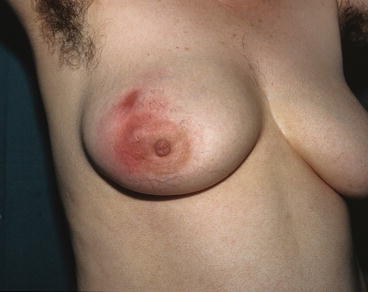

Fig. 8.2
Plurifocal lactating mastitis
Non–lactating mastitis (Fig. 8.3) is less common and rarely infectious and has a variable microbiology and may not resolve with antibiotic therapy.
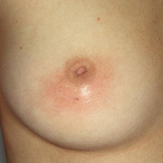

Fig. 8.3
Non-lactating mastitis (subareolar mastitis) associated to nipple inversion
Main characteristics of acute peripheral lactating abscess and acute subareolar non-lactating abscess are portrayed in Fig. 8.4.
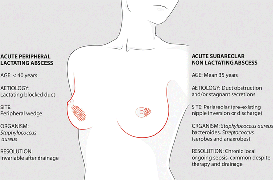

Fig. 8.4
Main characteristics of acute peripheral lactating abscess and acute subareolar non-lactating abscess
Surface mastitis mostly includes skin-related mastitis, a grouping of lesions common anywhere in the body and occasionally found in the breast. Actually, the simple term skin-related does not take into account breast inflammations that can originate from subcutaneous tissues as breast cellulitis, breast lymphangitis or breast oedema. With the term surface mastitis, we intend to encompass these superficial inflammatory lesions with skin-related lesions. In our opinion, Mondor disease too should be included in this group for its characteristic superficial presentation in the breast. Our classification for mastitis is shown in Table 8.1.
Table 8.1
Personal classification of mastitis
Lactating mastitis |
Non-infectious (engorgement) |
Infectious simple |
Infectious complicated (abscess) |
Non–lactating mastitis |
Neonatal and pubertal |
Subareolar mastitis |
Peripheral mastitis |
Postsurgical infections |
Mastitis associated with malignancy |
Surface mastitis |
Skin-related infections |
Breast cellulitis |
Breast lymphangitis |
Breast oedema |
Mondor disease |
History. In the presence of mastitis, the diagnosis is based primarily on the history and clinical symptoms. The general history should investigate generic factors of risk for infection as diabetes, chronic diseases, HIV infection, or an impaired immune system. Since almost all kinds of mastitis tend to recur, an anamnestic record of prior episodes is equally important.
Clinical presentation. A breast infection is usually diagnosed based on physical examination. Features may be various. In general, a painful, swollen lump in the breast is found, sometimes with redness, heat and swelling of the overlying skin. Fever and malaise may or may not be present according to the nature of infections, but symptoms could subside in case the woman took antibiotics early.
Assessment. In general, an ultrasound may be useful to determine the liquid or solid nature of the lesion and guide a fine needle aspiration, if appropriate. Mammography in the acute phase is of little use.
TREATMENT OF INFECTIOUS MASTITIS (principles) – Classification between epidemic and sporadic (or endemic) mastitis is still used but has given way to recognition that these infections form a spectrum of illnesses, depending upon the virulence of the infecting organism and the degree of bacterial colonisation of the milk.
Epidemic mastitis is a hospital-acquired infection caused by virulent strains of Staphylococcus aureus. This infection is rare, and it usually occurs within 4 days of delivery. Even with prompt antibiotic therapy, progression to abscess formation may occur.
Non–epidemic (or sporadic) mastitis, in contrast, is a milder infection with less virulent organisms and generally responds well to treatment without hospitalisation. Hospitalisation may be required, however, if the infection fails to respond. Keeping the breast empty of milk promotes healing by helping to drain the culture medium that facilitates growth of organisms. Hence, the earlier recommendations that breastfeeding cease while mastitis is being treated have been superseded by the knowledge that breastfeeding is generally not harmful to the infant – when using appropriate antibiotics – and may speed resolution of the infectious process.
Antibiotics are effective in lactating mastitis. In mastitis outside of breastfeeding, antibiotics may be disputable and, if indicated, should cover the spectrum of both aerobic and anaerobic bacteria, be administered for not less than 15 days and sometimes extended up to 2 months.
In the presence of an infection that does not respond to treatment, more investigations should exclude the presence of an underlying pathology as a BC.
TREATMENT OF BREAST ABSCESS (principles) – Any abscess should be managed by aspiration or incision and drainage preceded and followed by a course of antibiotics. In deep abscess, aspiration should be preferred, while in superficial abscess, incision is recommended. If the skin overlying the abscess is suffering, then the impaired skin should be excised [1].
Aspiration is best performed with ultrasound guidance with the abscess cavity being washed with local anaesthetic to dilute out and to help aspirate pus and reduce pain (Fig. 8.5). Repeated aspiration two to three times every 2–3 days is required to achieve resolution of most breast abscesses. Only for larger abscesses aspiration may need to be repeated five or more times.
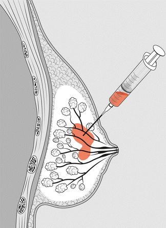

Fig. 8.5
Needle aspiration of a deep breast abscess in order to evacuate pus and wash the cavity with local anaesthetic
Incision and drainage, if indicated, can almost always be performed under local anaesthesia or slight sedation. Only a small incision is required to drain a breast abscess adequately (Fig. 8.6) when feasible. An incision is made usually at the lower edge of the abscess to allow the pus out and ensure that it will continue to drain when the patient is sitting upright in the post-operative period. After incision, the abscess is irrigated with the same local anaesthetic solution to wash out residual pus and to limit the pain of the procedure. Placement of a drain or packing the abscess cavity after incision and drainage is usually unnecessary.
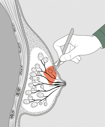

Fig. 8.6
Drainage of a superficial breast abscess with a small incision
However, if a large abscess is present, surgical drainage may require general anaesthesia. In this case, the wound is packed initially with a wick soaked in antiseptic such as iodine solution and will not normally be closed with sutures. This helps to better clean the wound and prevent bleeding. It also ensures that the wound stays open to allow any remaining infection to drain out. If the skin is closed immediately or soon after surgery, there would be a high chance that the infection would persist and the abscess would recur. For this reason, it is wise to let the wound close in its own time.
WORKUP – In conclusion, if an infectious mastitis is suspected:
Treat as soon as clinically suspected.
Re-evaluate every 3 days until completely cleared.
Change the antibiotic prescription if no response after 1 week.
Consider minimal biopsy (skin punch) if not responding to second antibiotic therapy.
Culture is usually not indicated.
If an abscess is suspected:
Aspirate and drain with local anaesthesia under ultrasound guidance.
A large (14–18 gauge) cannula needle is advisable.
Following ultrasound could confirm the presence of small or residual abscess.
Re-evaluate every 3 days until completely cleared.
Treat with antibiotic if there are systemic symptoms (fever) or local cellulitis.
Cultures are not necessary, unless there is no response to treatment.
Minor surgical incision for drainage is reserved to very superficial abscess
Major surgical drainage is rarely necessary.
A complete overview on mastitis, aimed to bring together available information on lactational mastitis and related conditions as well as their causes, is published by World Health Organization (see Useful Websites at the end of this chapter).
8.2 Lactating Mastitis
Clinical Practice Points
Lactating mastitis is the most common form of infectious mastitis, even if a real infection during breastfeeding is less common than believed to be.
Early prescription of appropriate antibiotics in case of true infection limits abscess formation.
In lactating mastitis, drainage may be a minor procedure performed by needle aspiration or minimal surgical incision.
Delay in referral to breast clinics of patients with lactating infection that does not settle rapidly with antibiotics continues to be a problem.
8.2.1 Non-infectious Lactating Mastitis
Non-infectious lactating mastitis is an engorgement of mammary ducts that can occur during breastfeeding as well as after weaning.
Breastfeeding engorgement is more common than infectious mastitis. Symptoms of simple engorgements are painful breast; tender, red, swollen and hard area of the breast, usually in a wedge-shaped distribution; slight or no fever; and no general malaise.
Milk removal and local packs may help to solve the symptoms, while investigations are not routinely required. For milk removal, breastfeeding is often more effective in pain control than using a breast pump and is more influential at encouraging milk flow. The pain of lactation mastitis is relieved by the application of gel packs or cold cabbage leaves to the breast, both are equally effective. Also, paracetamol may give some relief. Long-lasting milk removal and better maternal and infant hygiene may reduce the recurrence.
Weaning engorgement. Breast engorgement may occur also after weaning. The pregnancy-/lactation-related hormones usually return to normal levels shortly after weaning, but for some women, it can take several months to rebuild non-lactating state and there is an increased risk of rebound lactation and mastitis before hormone levels settling.
Most cases of postweaning mastitis or breast engorgement are solved with relatively little treatment. Recurrent postweaning mastitis on the other hand can be an indicator of a developing hyperprolactinemia or thyroid disorders and endocrinological examination must be considered.
Cold wraps or also warm, lactation-inhibiting herbs or medications can be used. Avoiding stress is equally important. Salvia officinalis and chasteberry extract can improve prolactin levels, which may reduce risk of recurrence, but no established data are available for its use in weaning engorgement. Bromocriptine may be effective in the prescribed dose but this rarely justifies the unpleasant side effects. Other prolactin-lowering medications (cabergoline, lisuride) are effective and appear safe but should be cautiously used for weaning.
8.2.2 Infectious Lactating Mastitis
Infectious lactating mastitis is rather common during breastfeeding, but less than believed to be. It is more frequent following a first child and most commonly seen within the first 6 weeks of breastfeeding, although some women develop it later or during weaning. Some blocked ducts could reduce drainage of milk from the affected area [2]. Moreover, there is usually a history of a cracked nipple or skin abrasion, although this is not always the site of entry of organisms. Staphylococcus aureus is the most common organism responsible, but Staphylococcus epidermidis and Streptococci are occasionally isolated.
Clinical presentation. The symptoms of lactating infection can develop more or less quickly. Most women first experience flu-like symptoms and just after a while, they may notice a sore red area on the breast, an abnormal discharge from the nipples or a prolonged, unexplained breast pain. Increasing pain is followed by other symptoms, as erythema, swelling and induration of the breast, associated with general symptoms of infection including nausea, fever 38 °C or higher, shivering and chills, fatigue and tachycardia. Some women do experience Raynaud’s of the nipple during breastfeeding, which can cause more considerable pain.
Treatment. If symptoms do not improve, or are worsening after 12–24 h despite effective milk removal, and there is severe deep burning breast pain, a ductal infection should be highly suspected.
Promotion of milk drainage and early antibiotic therapy are the cornerstones of treatment. A 10-day course of a penicillinase-resistant antibiotic such as flucloxacillin (or erythromycin if penicillin allergic) is required. Tetracycline, ciprofloxacin and chloramphenicol should not be used to treat lactating breast infection as they may enter breast milk and can harm the baby.
Antibiotics give satisfactory resolution in many cases. If the infection does not settle after one course of antibiotics, if no pus is detected on ultrasonography and if clinical and imaging assessments indicate that the lesion is actually infective, antibiotics should be changed to cover other possible pathogens.
The role of associated mycosis and the value of fluconazole in breast pain and infection associated with breastfeeding are controversial. The evidence that fungal infections are important causes of mastitis is largely anecdotal. There are no data from properly controlled clinical studies showing the value of fluconazole and it should not be prescribed until further clinical trial evidence shows it to be beneficial.
Frequent emptying of both breasts by breastfeeding is essential. Also essential is adequate fluid supply for the mother and the baby. Use of pumps to empty the breast is somewhat controversial and anyway not as efficient as breastfeeding. In cases of minor mastitis, massage and application of heat prior to feeding can help as this may help unblock the ducts. However, in more severe cases of mastitis, heat or massage could make the symptoms worse and cold compresses are better suited to contain the inflammation.
At this point, investigations are not routinely required. Culture of the breast milk is proposed only when:
Antibiotics have been prescribed and there has been no response after 48 h.
Mastitis is severe enough also before any antibiotics are prescribed.
Mastitis is recurrent.
Infection is likely hospital acquired (endemic).
The woman is unable to take standard antibiotics (such as flucloxacillin and erythromycin).
Breast abscess develops only rarely, in about 0.5 % of breastfeeding women. Known risk factors are age over 30, primiparous and late delivery. No correlation was found with smoking status; however, this may be in part because much fewer smoking women choose to breastfeed. If a breast abscess is suspected, or needs to be excluded, an initial ultrasound is useful in assessing the presence of collection of fluids.
An established abscess, usually in the middle or in the peripheral part of the breast tissue, should be treated either by repeated aspiration every 2–3 days until no more pus is aspirated or by incision and drainage. With ultrasound-controlled aspiration, minor fluid collection can also be evacuated. Surgery should be reserved for the minority of cases that do not resolve with antibiotics and repeated aspiration or those where the abscess is superficial with an involvement of the overlying skin (see Sect. 8.1 above).
Breast infections in lactating women can be distressing and debilitating and can interfere with early baby bonding. Women who want to continue breastfeeding should be encouraged to feed with the unaffected breast and, once letdown occurs in the affected breast, feed with the affected breast until it is completely empty. Warm compresses and paracetamol may give some relief.
For those women who present with multiple areas of breast infection, and are exhausted by breastfeeding, a consideration should be given to stopping breastfeeding and halting milk flow. Stopping milk production is achieved by prescribing cabergoline 2.5 mcg given twice a day for 2 days.
8.3 Non-lactating Mastitis
Clinical Practice Points
As in lactating infection, non-lactating mastitis may be non-infectious or infectious without or with abscess formation.
Non-lactating mastitis can be separated into those that occur centrally in the subareolar region and those that affect the peripheral breast tissue.
Subareolar breast abscesses are slightly trickier. In subareolar periductal mastitis, cigarette smoking is a powerful facilitator of severe inflammatory complications.
In mastitis outside of breastfeeding, the administration of antibiotics, which cover the spectrum of both aerobic and anaerobic, must not be less than 10–15 days, also being able to extend up to 2 months.
In the presence of an infection that does not respond to treatment, it is good to do investigations to exclude the presence of a BC.
Besides neonatal and pubertal mastitis, non-lactating mastitis falls into two main groups:
Subareolar mastitis, related to periductal inflammation, independent or related to duct ectasia
Peripheral mastitis, sometimes related to systemic diseases, as diabetes and rheumatoid arthritis, or infections, as tuberculosis.
8.3.1 Neonatal and Pubertal Mastitis
Neonatal breast infection is not common but can occur in the first few weeks of life when the breast bud is enlarged (see Sect. 3.4). Although Staphylococcus aureus is the usual organism, occasionally, infection is due to Escherichia coli. If an abscess develops, a small incision placed as peripherally as possible to avoid damaging the breast bud leads to rapid resolution [3].
Prepubertal girls may also develop breast abscesses and this topic is treated in Chap. 3. Also as in neonatal infection, the decision for surgical drainage should be carefully made because future breast deformation may occur.
8.3.2 Subareolar Mastitis
Stay updated, free articles. Join our Telegram channel

Full access? Get Clinical Tree


