Fig. 31.1
(a–c) Preoperative view. The scars from prior quadrantectomies are in the upper outer quadrant of both breasts. Dots indicate the microcalcifications. Preoperative drawings for inferior pedicle reduction mammoplasty
31.2 Surgery
Bilateral quadrantectomy was done under stereotactic wire guidance, and the entire upper outer quadrants were resected. The defect was reconstructed with an inferior pedicle. Sentinel lymph node biopsy found one negative lymph node in the right and two negative nodes in the left breast.
31.3 Clinical and Cosmetic Outcome
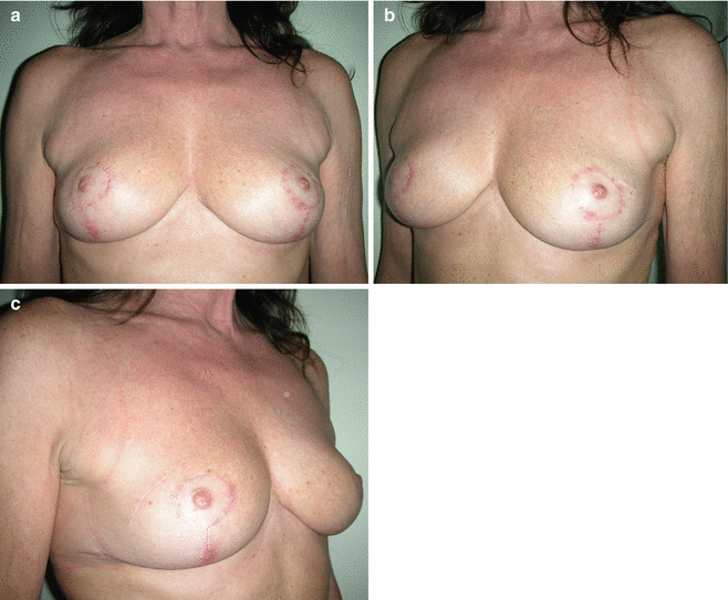
The postoperative course was uneventful. Histological examination showed high-grade DCIS in both breasts and was completely resected. Radiation therapy was given for 6 weeks. Postoperatively the patient revealed adequate breast symmetry but with volume lacking in both the upper outer quadrants (Figs. 31.2a–c and 31.3).


Get Clinical Tree app for offline access

Fig. 31.2
(a–c) Postoperative view 3 years after surgery and radiation. Both breasts show adequate volume and symmetry but lack some volume in the upper outer quadrants

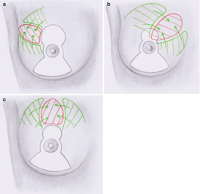
Fig. 31.3
(a–c) A large defect in the upper breast after quadrantectomy may be reconstructed with tissue from the medial quadrant (a


Stay updated, free articles. Join our Telegram channel

Full access? Get Clinical Tree


