■ WHIM syndrome: autosomal dominant, 1° immunodeficiency caused by a CXCR4 mutation – warts, hypogammaglobinemia, infections (bacterial), and neutropenia (2° to myelokathexis)
■ WILD syndrome: warts, immunodeficiency, lymphedema, and dysplasia (anogenital)
■ Treatments: destructive (cryotherapy, ED&C, scissors/shave removal, laser (PDL or CO2)/PDT, cantharidin, and salicylic acid preparations), immunomodulatory/antiviral (SADE/DPCP and intralesional immunotherapy [e.g., Candida]), and 5-FU (topically w/ salicylic acid usually or intralesional), intralesional (bleomycin and cidofovir gel)
• Mucosal/Genital manifestations of HPV infection
■ Genital warts (condyloma acuminata)
○ Occur on external genitals/perineum/perianal/groin/mons/vagina/urethra/anal canal
○ Smooth, sessile, raised, skin-colored to brown lobulated papules
○ HPV-6, HPV-11, HPV-16, HPV-18, HPV-31, HPV-33, and HPV-45
○ Condylomata plana (flat cervical warts) best seen w/ acetic acid → whitening
○ Most cases resolve spontaneously within 2 years
○ RFs: sexual intercourse at young age, # of sexual partners, and MSM
○ Circumcision →↓risk HPV transmission
♦ Most common scenario = persistent cervical infection with high-risk HPV type (HPV-16, HPV-18, HPV-31, HPV-33, and HPV-45)
♦ Immunosuppression (e.g., HIV+) can ↑risk
○ Histology: epidermal hyperplasia, koilocytosis (should be seen in stratum spinosum too), papillomatosis (less severe and more rounded than in common warts), and parakeratosis
○ Treatments: destructive (cryotherapy, TCA (higher concentrations), electrosurgery, scissors/shave removal, laser (CO2)/PDT, and podophyllotoxin/podophyllin), immunomodulatory/antiviral (imiquimod, sinecatechins, intralesional immunotherapy, and cidofovir gel/intralesional)
○ HPV vaccines: contain L1 major capsid protein (self-assembles into virus-like particles → allow for development of immunity without any harm because they do not contain DNA)
♦ Three types: quadrivalent (Gardasil; HPV-6, HPV-11, HPV-16, and HPV-18), bivalent (Cervarix; HPV-16 and HPV-18), and 9-valent (HPV-6, -11, -16, -18, -31, -33, -45, -52, and -58)
♦ Best to use before sexually active – FDA-approved for females and males 9 to 25/26 years old
■ Bowenoid papulosis: multiple brown papules/smooth plaques on genitals/perineum/perianal that are high-grade squamous intraepithelial lesions (HSIL) or SCCIS; progression to invasive SCC is very rare; a/w high-risk HPV types
■ Erythroplasia of Queyrat: red smooth plaque on glabrous penis/vulva that is HSIL or SCCIS; increased risk of progression to invasive SCC; has high-risk HPV types
■ Buschke-Lowenstein tumor (arises on genitals)
○ Part of a group of verrucous carcinomas (slow growing and locally destructive) that includes oral florid papillomatosis (HPV-6, HPV-11; RFs: smoking, radiation, and inflammation), epithelioma cuniculatum (HPV-2, HPV-11, and HPV-16), and papillomatis cutis carcinoides
○ Cauliflower-like tumors that infiltrate deeply on external genitals and perianally
○ Histology: papillomatous acanthotic epidermis with bulbous (“pushing”) downward-extending rete ridges; no cellular atypia/basement membrane penetration
○ Treatment: excision with clear margins
■ Oral warts: soft pink-white papules on any oral surface; HPV-6 and HPV-11; more common in HIV
○ Focal epithelial hyperplasia (Heck’s disease): multiple flat wart-like papules on gingival/buccal/labial mucosa in children (esp. South American); HPV-13 and HPV-32
■ Recurrent respiratory papillomatosis: papillomas of airways due to HPV-6 and HPV-11; #1 benign tumor of larynx; hoarseness + stridor + respiratory distress; childhood (2° to vertical transmission) and adulthood (2° to genital-to-oral contact) onsets; can → SCC, esp. in smokers
II Human herpes viruses
• A total of 8 distinct human herpesviruses (HHV-1 to HHV-8) belong to the Herpesviradae family; all are characterized by an icosahedral capsid containing linear double-stranded DNA, surrounded by a glycoprotein-containing envelope; replicate in host nucleus
• Pathogenesis involves infection, latency, and reactivation
Herpes simplex virus (HHV-1/HSV-1 and HHV-2/HSV-2)
• Recurrent vesicular eruptions occurring in orolabial (classically HSV-1) and genital (classically HSV-2) regions
• Primary infection = first infection with virus (may → symptoms); latency = virus lies dormant in sensory (dorsal root) ganglia; reactivation/recurrence (may → symptoms)
• Genital herpes RFs: 15 to 30 years old, ↑sexual partners, lower income/education, HIV(+) (vice versa too – genital HSV-2 → ↑HIV risk), and homosexuality
■ Infection can occur without clinical lesions (and often does), and virus may still be shed
■ HSV-1 spread by saliva/secretions and HSV-2 spread by sexual contact → viral replication at skin/mucous membrane → retrograde axonal flow to dorsal root ganglia → latency and subsequent reactivation
■ HSV can evade host immune system (e.g., ↓expression of CD1a by APCs, ↓TLR signaling)
■ Reactivation triggers: stress, UV (UVB > UVA), fever, injury (e.g., chemical peel or fractionated laser), and immunosuppression
■ Classic appearance: grouped/clustered vesicles on a red base
○ Can become pustules, erosions (with classic scalloped borders due to coalescence), and ulcers, ultimately crusting over and healing within 6 weeks
■ 1° infection: 3 to 7 days postinfection → prodromal symptoms (tender lymphadenopathy, malaise, anorexia, and fever) → mucocutaneous lesions +/− pain/tenderness/burning/tingling just before lesions erupt
■ Recurrent infections: generally milder than 1° infections, have 24 hour prodrome of tingling/itch/burning
○ 1° HSV can be severe (gingivostomatitis in children; pharyngitis/mononucleosis-like in adults)
○ Mouth (esp. buccal mucosa and gingivae; favors anterior mouth unlike herpangina) and lips (recurrent lesions prefer vermilion border) affected
○ 1° infection often asymptomatic, but can → painful/tender erosions on external genitalia, vagina, cervix, buttocks, and perineum (women) +/− lymphadenopathy/dysuria (women mainly)
♦ 1° worse in women – ↑% extragenital involvement, urinary retention, and aseptic meningitis (10%)
○ Recurrent – mildly symptomatic with few vesicles lasting about 1 week; frequency of outbreaks usually decreases over time
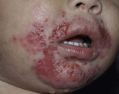
○ Eczema herpeticum: widespread, sometimes severe HSV infection in areas of atopic dermatitis, Hailey-Hailey, or Darier’s disease (Fig. 5-2) +/− systemic symptoms, lymphadenopathy, may be life-threatening; ↑in children
♦ Usually HSV-1; associated with Th2 shift in immune system
♦ ↑in patients with severe atopic dermatitis w/ onset <5 years old, ↑IgE levels, ↑eosinophils, and food/environmental allergies
♦ Have been associated with topical calcineurin inhibitors
○ Herpetic whitlow: infection of digits (HSV-1 in children and HSV-2 in adults) w/ vesiculation/pain/swelling; recurrence seen; bimodal peaks at <10 years old and 20 to 40 years old
○ Herpes gladiatorum: HSV-1 infection 2° to athletic contact (classically on lateral neck/side of face and forearm)
○ HSV folliculitis (herpetic sycosis): follicle-based vesicles/pustules in beard-area (HSV-1)
○ Severe/chronic HSV: large, chronic ulcers may involve respiratory or GI tract; more common in immunocompromised
○ Ocular HSV: keratoconjunctivitis w/ lymphadenopathy and branching dendritic corneal ulcer; blindness may occur (HSV-2 in newborns; HSV-1 otherwise)
○ HSV encephalitis: most common fatal viral encephalitis in the United States (>70% die without tx); can be associated with mutations in TLR-3 or UNC-93B; usually HSV-1; fever/altered mentation/strange behavior; temporal lobe #1 site
○ Neonatal HSV– see Pediatric Dermatology section
■ Viral culture (high specificity, low sensitivity), direct fluorescent antibody assays, serology (Western blot = gold standard), PCR (most sensitive/specific), and Tzanck smear (multinucleated epithelial giant cells; best when done on acute lesions)
■ Histology: intraepidermal vesicle + slate-gray enlarged keratinocytes (ballooning degeneration) which are multinucleated with margination of chromatin
○ +/− Cowdry A inclusions (eosinophilic inclusion bodies) within nucleus, epidermal necrosis, multicellular dermal infiltrate, and perivascular cuffing
■ Orolabial: oral penciclovir/valacyclovir, topical penciclovir, or topical acyclovir/hydrocortisone combination
■ Genital: oral acyclovir/famciclovir/valacyclovir
○ Use meds w/in first 48 hours → ↓pain/healing time/viral shedding
○ Suppressive daily doses may be given in patients with >6 outbreaks of orolabial/genital HSV per year (also ↓viral shedding)
■ May need IV acyclovir in eczema herpeticum, neonatal HSV, or severe HSV in immunosuppressed
■ Foscarnet or cidofovir for acyclovir-resistant HSV (more common in immunosuppressed patients)
• Boards factoid: HSV-1 is the most common cause of EM minor (herpes associated EM; HAEM)
Varicella zoster virus (HHV-3)
• Causes varicella (chickenpox) and herpes zoster (shingles)
• Varicella is the 1° infection and herpes zoster is the reactivation of the latent infection (more common in immunosuppressed and elderly and can → death, e.g., via SIADH development in disseminated zoster patients)
• Primary varicella incidence has decreased because of VZV vaccination
• Herpes zoster occurs in 20% of adults, 50% of immunocompromised
■ RFs: physical and emotional stress, fever, trauma, and immunosuppression
■ Transmitted via aerosolized droplets and direct contact with lesional fluid
○ Contagious from 1 to 2 days before lesion develops in varicella until all lesions crusted over
■ After primary varicella infection, VZV travels to dorsal root ganglion and stays dormant – if reactivated later will replicate, travel down sensory nerve to the skin, and present as herpes zoster
○ Primarily self-limited in healthy individuals
♦ More severe disease in adolescents and adults
○ Prodromal symptoms: fever, fatigue, and myalgias
○ Cephalocaudal progression of classic lesions described as “dew drops on rose petal:” vesicles on an erythematous base that become pustular, then crust over
♦ Crops of lesions in various stages
○ Vaccine-associated varicella zoster may rarely develop after the vaccine is administered – represents mild case of chickenpox that may start at injection site
○ Primary varicella in pregnancy
♦ Congenital varicella syndrome: cutaneous scarring; CNS/ocular/limb anomalies; risk greatest if infection occurs during first 20 weeks of gestation; exposed fetus may develop reactivation (herpes zoster) in childhood
♦ Neonatal varicella: perinatal varicella transmission (within 5 days before delivery until 2 days postdelivery); disease is severe (up to 30% mortality) because of the lack of protective maternal antibodies
■ Herpes zoster: prodrome (itch, tingling, hyperesthesia, and pain) → painful grouped vesicles on red base in a dermatomal pattern
○ Trunk = most common location (thoracic); face #2 (cranial; trigeminal nerve most common nerve involved); lumbar #3, and sacral #4
○ Postherpetic neuralgia: pain, potentially chronic, after lesions have cleared; more common, severe and chronic in elderly
○ In HIV patients, lesions more persistent and thickened
○ Disseminated disease = dermatomal disease + >20 lesions outside of dermatome +/− visceral involvement; almost exclusively seen in immunosuppressed (AIDS, lymphoreticular malignancy, long-term immunosuppressive medication use, etc.); increased risk of life-threatening pneumonitis and encephalitis
○ Vasculopathies (usually of CNS, but also peripheral arteries) are a worrisome delayed complication
○ Dermatomal-specific herpes zoster findings:
♦ Ramsay-Hunt syndrome: disease of geniculate ganglion of facial nerve (CN-VII) may → ear pain, vesicles on tympanic membrane and EAM; ipsilateral facial nerve paralysis, dry mouth/eyes, anterior 2/3 tongue taste loss, and auditory (e.g., deafness and tinnitus) and equilibrium issues (vestibulocochlear nerve)
♦ Aseptic meningitis and/or vasculopathy (encephalitis) if CN-V affected
♦ Hearing impairment/deafness if CN-VIII affected
♦ Eye involvement (herpes zoster ophthalmicus) if CN-II, CN-III, or CN-V affected
➔ Hutchinson’s sign (involvement of the side and tip of nose): indicates disease of the external division of the V1 nasociliary branch; may → to ocular involvement (e.g., keratitis, uveitis, acute retinal necrosis, and visual loss) 3/4 of time
➔ Uveitis is most common form of ocular involvement; keratitis #2
♦ Bell’s palsy if CN-VII affected
♦ Back dermatome complications
➔ Cervical: motor neuropathy of arm (with possible atrophy) and diaphragm weakness
➔ Thoracic: abdominal wall pseudohernia and weakness of muscles
➔ Lumbar: motor neuropathy of leg (with possible atrophy)
♦ Possible urinary hesitancy/retention if sacral dermatomes involved
♦ Possible dilatation, constipation, pseudo-obstruction, reduced anal sphincter tone w/ thoracic/lumbar/sacral zoster
• Diagnosis: Tzanck smear, DFA, PCR (sensitive, fast), viral culture (specific, not sensitive), serology (four-fold increase in IgG titer can retrospectively confirm prior infection), and skin biopsy (similar appearance to HSV, but immunohistochemistry can differentiate)
○ Treatment with systemic acyclovir or valacyclovir within 3 days of lesion onset → ↓severity/duration disease
♦ Oral administration appropriate in healthy children/adults
♦ IV acyclovir in immunocompromised patients
♦ Varicella vaccine may be given within 72 to 120 hours of exposure in nonimmune, immunocompetent individuals >12 months
♦ VZIg (Varicella zoster immunoglobulin) should be administered within 96 hours of exposure in immunocompromised, pregnant females, and neonates
➔ IVIg may alternatively be administered
♦ Oral acyclovir can be administered within 7 to 10 days of exposure
○ Primary prevention = varicella vaccination
♦ Live attenuated virus recommended as a 2 dose vaccination series; part of primary immunization series
♦ Initial dose at 12 to 15 months, booster dose at 4 to 6 years
♦ Contraindicated in pregnancy and in immunocompromised patients
○ Sequelae of primary varicella
♦ Reye’s syndrome in setting of aspirin administration (now rare)
♦ Pneumonia more common in older individuals; high mortality if untreated
♦ Encephalitis, cerebella ataxia, and hepatitis
○ Antiviral treatment with acyclovir (IV form in immunosuppressed), famciclovir, or valacyclovir is best given within 72 hours; prednisone helps with acute pain but has no effect on course or development of PHN
♦ ↓rate of postherpetic neuralgia (PHN) in patients >50 years old
♦ Valacyclovir and famciclovir preferable to acyclovir
○ PHN: tricyclic antidepressants (e.g., nortriptyline), gabapentin, 8% capsaicin patch, pregabalin, opioid analgesics, and lidocaine patch
○ Live attenuated vaccine → ≈50%↓ in development of disease and 67%↓ in PHN; for immunocompetent patients >60 years old
Epstein-Barr virus (HHV-4)
• Causes infectious mononucleosis plus many other disorders (e.g., oral hairy leukoplakia, hydroa vacciniforme, Gianotti-Crosti syndrome, genital ulcers, and various hematologic disorders/malignancies (e.g., Burkitt’s lymphoma, NK/T-cell lymphoma, post-transplant lymphoproliferative disorder, and nasopharyngeal carcinoma)
• Pathogenesis: transmission via saliva/blood → infects mucosal epithelial cells initially → B-cells (where virus can lay dormant and evade immune system via production of EBNA-1 protein and latent membrane protein-2)
■ Incubation period of 1 to 2 months; symptoms develop with viral replication
■ In patients with ↓cell-mediated immunity, infected B-cells may continue to replicate → lymphoproliferative disorders (cell-mediated immunity appears to be more important than humoral, conferring immunity after first mononucleosis episode)
■ Mononucleosis: typically young adults w/ pharyngitis, fever, and cervical lymphadenopathy
○ Splenomegaly (and possible rupture) +/− hepatomegaly
○ Lymphocytosis (up to 40% atypical lymphocytes)
○ May have nondistinct polymorphous (e.g., urticarial, morbilliform) eruption in 5% to 10% occurring within first week of illness
♦ Petechial lesions on eyelid and hard/soft palate junction
♦ +/− genital ulcers (esp. females)
○ Ampicillin/amoxicillin → “hypersensitivity” skin reaction (itchy generalized morbilliform eruption → desquamation)
■ Oral hairy leukoplakia: corrugated white plaque typically on lateral tongue, with strong HIV association; more common in smokers
■ Gianotti-Crosti syndrome and papular-purpuric glove and stocking syndrome (more common w/ parvovirus B19, though) may occur in setting of EBV infection
■ Monospot test: nonspecific, confirms presence of IgM heterophilic antibodies which are often present in EBV infection and may persist for months after infection; 85% of older children/adults are positive during second week of infection, but Monospot is often negative in younger children
■ EBV-specific antibodies: higher sensitivity in younger children; can be useful in determining current vs prior infection (Table 5-1)
○ VCA (viral capsid antigen) IgM/IgG, EA (early antigen) IgG, and EBNA IgG
Table 5-1
Epstein–Barr Virus-Specific Serology Interpretation
| Viral Capsid Antigen (VCA) | ||||
| Status | IgG | IgM | EA | EBNA |
| No past infection | − | − | − | − |
| Acute IM | + | + | ± | − |
| Convalescent IM | + | ± | ± | ± |
| Past infection | + | − | Low + or − | + |
| Reactivated/chronic | ++ | ± | ++ | ± |
■ CBC may reveal lymphocytosis with atypical lymphocytes and thrombocytopenia
■ Transaminitis may be present
■ Positive heterophilic antibody (>1 : 40) and >10% atypical lymphocytes suggests acute infection
■ PCR to EBV DNA may be performed from tissue or blood; RT-PCR available from lymphoid cells
■ Oral corticosteroids may be considered for severe cases of tonsillitis
■ Avoid contact sports until splenomegaly resolves (risk for splenic rupture)
■ Rare sequelae: upper airway obstruction, aseptic meningitis, meningoencephalopathy, myocarditis, pericarditis, and renal failure
Cytomegalovirus (HHV-5)
• Transmitted via body fluids, fomites, vertical transmission, transplanted organs, and hematopoietic stem cells
• Infects leukocytes → dissemination → various organs → latency
■ Most infections are asymptomatic in healthy adults; however, can cause severe disease in utero (TORCH), or in immunosuppressed/transplant patients (CMV retinitis/blindness, meningoencephalitis, pneumonitis, GI ulcers)
■ After the 1° infection, very low risk of reactivation, except for immunocompromised patients
• Cutaneous features in adults
■ Mononucleosis-like presentation (e.g., sore throat, fever, lymphadenopathy, and hepatosplenomegaly) may be associated with nonspecific exanthem (e.g., morbilliform)
○ If ampicillin given → eruption (as in infectious mononucleosis)
■ Recalcitrant ulcers of perineum or leg in HIV patients; these patients may also get verrucous plaques, vesicles, and/or nodules
• Diagnosis via human fibroblast culture (gold standard), but faster methods include shell vial assay, PCR, and serologic testing; histology of ulcers may show enlargement of endothelial cells with pathognomonic “owl’s eye” (intranuclear) inclusions
• Ganciclovir (IV) and valganciclovir (oral) are first-line treatments
HHV-6 (Roseola infantum, exanthem subitum, sixth disease)
• One of the most common viral exanthems of childhood (discussed in detail in Pediatric Dermatology chapter); up to 15% of infants may develop febrile seizures, but otherwise follows a generally benign course in healthy pts
■ 95% of pts are between 6 months to 3 years of age
• Virus remains latent in T cells for life → reactivation has been a/w pityriasis rosea (along with HHV-7) and DRESS syndrome (along with EBV, CMV and HHV-7)
HHV-7
• Lymphotropic virus that shares significant homology with HHV-6 and may participate in co-infection w/HHV-6
• Although not definitively causative of any disease, it has been a/w pityriasis rosea (along with HHV-6), and a subset of exanthem subitum cases (co-infection with HHV-6; unique clinical presentation)
III Other viruses not covered elsewhere
Poxviruses
• Smallpox (Variola virus; Orthopox genus)
■ Infection via respiratory tract → 7 to 17 days incubation period → 1 to 4 days prodromal period (fever, headache, myalgias, and malaise) → centrifugal (face/arms/legs > trunk) vesiculopustular eruption and may involve hands/feet (lesions in any given anatomic region will be in same stage) w/ lethargic/“toxic” appearance
○ Rash: macule → papule → vesicle → pustules; typically scarring
○ Lesions first appear on palms/soles
○ Patients infectious from eruption onset till 7 to 10 days posteruption
○ Oral lesions (tongue, mouth, and oropharynx) often appear before cutaneous by lesions 1 day
■ Complications: blindness, encephalitis, toxemia, hypotension, pneumonitis, arthritis, and osteitis
■ Diagnosis: PCR, viral culture
■ Treatment: supportive; vaccine as prophylaxis
• Vaccinia (Vaccinia virus; genus = Orthopox): used for live smallpox vaccine
■ SEs: lymphadenopathy, ocular vaccinia, generalized vaccinia, vesiculopustular/urticarial/morbilliform eruption, eczema vaccinatum (in patients with atopic dermatitis, Darier’s, or Hailey-Hailey disease), erythema multiforme, postvaccinial CNS disease, and progressive vaccinia (immunosuppressed patients; can → death)
• Monkeypox (Monkeypox virus; genus = Orthopox): central/western Africa, though United States outbreak from prairie dogs
■ Can spread via cutaneous inoculation or inhalation (hosts are monkeys, rodents, or humans)
■ Prodrome (fever/sweating/chills) → smallpox-like lesions, but usually milder/fewer lesions
○ Lesions may present in various stages and favor face and extremities (esp. palms/soles), with centrifugal spread; may scar
○ May have systemic symptoms (respiratory, fever, and LAD in 67%)
• Cowpox (Cowpox virus; genus = Orthopox): Europe and Asia
■ Spread via cutaneous contact (hands and face) with infected animal (usually cats)
○ Incubates 7 days → painful red papule at contact site → vesicular → pustular → hemorrhagic → ulcer w/ eschar
○ Lesions usually solitary and occur on hands/fingers
• Orf (ecthyma contagiosum; Orf virus; genus = Parapox): as a result of contact with infected animals (sheep, goats, or reindeer; usually on udders/perioral areas of ewes)
■ Develop one to few lesions at contact site (usually hands)
■ RFs: certain jobs (shepherds, butchers, and veterinarians)
■ Six lesion stages: maculopapular (umbilicated) → targetoid → acute (weeping nodule) → regenerative (nodule w/ thin crust and black dots) → papillomatous → regressive (crust overlying resolving lesion)
■ Diagnosis via histology (depends on stage) or PCR
• Milker’s nodules (“Pseduocowpox;” Paravaccinia virus; genus = Parapox): papules at site of contact (usually muzzles of calves and teats of cows)
■ Distal upper extremities usually with single lesion(s), which look like orf
■ Most common in farmers/ranchers, veterinarians, and butchers
■ Diagnosis via histology or PCR
• Molluscum contagiosum (Molluscum contagiosum virus [MCV]; genus = Molluscipox)
■ Common infection in school-aged children; may be sexually transmitted in adolescents/adults
■ Cause by molluscipox infection
○ Two subtypes: MCV-I and MCV-II
■ Infection spread by contact with infected skin or fomites, or possibly via water
■ Prototypical lesion is an umbilicated, pink, and pearly papule
○ Most common distribution: intertriginous areas, torso, lower extremities, and buttocks
○ Lesions can become widespread in patients with impaired skin barrier (atopic dermatitis or ichthyosis) or immunodeficiency (chemotherapy-induced or HIV; may also see giant molluscum lesions)
■ Histology: molluscum bodies within dermis
■ Treatments: cryotherapy, cantharidin, extraction/curettage, cimetidine, candida antigen immunotherapy, topical retinoids, and imiquimod
■ Self-limited with resolution after weeks to years of infection
Chikungunya virus
• Single-stranded (+)sense RNA virus belonging to Togaviridae family; classified as an “arbovirus” because has arthropod vector
• Transmitted by Aedes (A. aegypti > A.albopictus) mosquitoes; endemic to Africa/India/Southeast Asia
• Symptoms: high fever, marked joint symptoms, (“Chikungunya” is an African word for “crooked/bent joints”) neuropathic acral findings, and headache/nausea/vomiting
• Cutaneous presentation: → morbilliform eruption (50%–75% of pts), mucosal aphthous-like ulcers, postinflammatory pigmentation of face/extremities, acral/facial edema, bullous eruptions in infants, and ecchymoses
Zika virus
• Icosahedral, single-stranded RNA virus within the Flaviviridae family
■ Flaviviridae family includes Yellow fever, Dengue fever, Japanese encephalitis, West Nile virus, and Zika virus
○ All are termed “arboviruses” because they are viruses that are transmitted by arthropods (mosquitoes or ticks most commonly)
○ Review the excellent JAAD 2016 CME article by Nawas et al, Emerging infectious diseases with cutaneous manifestations
• Most commonly transmitted via bites from infected Aedes aegypti and Aedes albopictus mosquitos
■ Virus may also be transmitted via blood transfusions, sexual contact, and most importantly, vertically (from mother to fetus) during pregnancy → microcephaly and other fetal anomalies
• In 2016, the WHO classified Zika as a global threat and the CDC raised issued their highest alert due to Zika’s association with microcephaly and possible a/w Guillain-Barré syndrome
■ No gender or age predilection
■ Incubation period of 3–12 days → 20% of infected adults develop mild symptoms lasting up to 1-2 weeks
○ Systemic symptoms: fever, myalgia, arthralgia, headache, and conjunctivitis
♦ Nonspecific, diffuse morbilliform/scarlatiniform eruption (begins 3 to 12 days after initial infection w/cephalocaudal progression; starts on face → spreads to trunk/extremities) → rash begins to subside after 3 days and completely resolves within 1 week of onset, sometimes with desquamation
♦ Mild hemorrhagic manifestations (petechiae and bleeding gums)
■ Unfortunately, there are no unique clinical features to differentiate Zika from other arbovirus infections → must consider dengue and chikungunya infection in your differential diagnosis
■ Confirmed with RT-PCR or ELISA during initial phase (first 7 days) of infection
○ Later in disease course, may check Zika-specific IgM antibodies and plaque reduction neutralization tests
■ Currently no vaccine exists, and no specific anti-viral therapies are available; avoid aspirin and NSAIDs (can worsen hemorrhagic sequelae)
○ People traveling to endemic areas should wear long-sleeved shirts and pants, stay in cool rooms that have screens on windows and doors, and use insect repellents (DEET); pregnant women should avoid travel to Zika-endemic areas!
Dengue virus
• Like Zika, West Nile virus and Yellow fever, Dengue is an arbovirus in the Flaviviridae family; also transmitted by Aedes aegypti mosquitoes
• Wide range of clinical presentations:
■ Asymptomatic infection: most common presentation (75% of cases)
■ Mild Dengue: very nonspecific, mimicking any other viral infection
■ Classic Dengue fever: fever, diffuse morbilliform/scarlatiniform rash (50% of cases), severe headache/myalgia/arthralgia, retroorbital pain, +/− petechial mucosal lesions, epistaxis and gingival bleeding
○ Classic Dengue fever rash: widespread erythema with characteristic white islands of sparing → heals with desquamation
■ Dengue hemorrhagic fever (DHF): more severe than classic Dengue fever; most likely to develop when a patient previously infected with 1 serotype is subsequently infected with a different viral serotype
○ Most common in children younger than 15yo
○ Symptoms: lethargy/weakness, vomiting, facial flushing, and circumoral cyanosis
■ Confirmed with RT-PCR or ELISA during initial/acute phase of disease, or IgM serologies later in disease course
■ Neutropenia helps distinguish from Chikungunya virus
• Treatment: No specific treatment; mainly supportive; avoid aspirin and NSAIDs (can worsen hemorrhagic sequelae)
Related posts:

Stay updated, free articles. Join our Telegram channel

Full access? Get Clinical Tree


