History and rationale for laser in the treatment of cardiovascular diseases
Early applications, growing expectations and the development of realistic perspectives
Current indications and applications: coronary, peripheral, TMR, EP
Technique of lasing and catheter technology
Adverse outcomes
Future potential
History
Lasers were first introduced to cardiovascular medicine in the 1980s. In light of the laser’s avid absorption in atherosclerotic material, the aim was to treat a variety of coronary and peripheral occlusions that were considered “non ideal” for standard balloon angioplasty.1 Initial studies predicted that laser would render coronary bypass surgery an unnecessary operation as preliminary experience reported successful plaque removal.2–4
Early Experience
Publication of several large scale successful clinical trials during early experience with laser led to a conviction that laser had established a prominent role in interventional cardiology.5,6 However, the application of the device in cardiac catheterization laboratories was fraught with technical difficulties. The devices were very large, cumbersome to handle and necessitated lengthy warm up and calibration time. The laser catheters were also very rigid and lasing technique with rapid advancement of the catheters across the target lesions was frequently utilized. This technique did not allow adequate absorption of the laser energy within the irradiated plaque. Consequently, laser induced spasm, thrombosis, dissections and perforations became a significant concern.7,8 Eventually, refinements in catheter design involving smaller laser generators9 and introduction of safe lasing techniques emphasizing slow debulking and concomitant injections of saline10,11 led to significant improvement in clinical outcomes.12 With better understanding of laser as a treatment modality, numerous indications were established for utilization within the field of cardiovascular medicine. At present, laser is used percutaneously in the cardiac catheterization suite for treatment of coronary and peripheral atherosclerotic vascular disease, and surgically for Trans-myocardial revascularization and for pacemaker/AICD lead removal in electrophysiology laboratories.
Current Indications and Applications
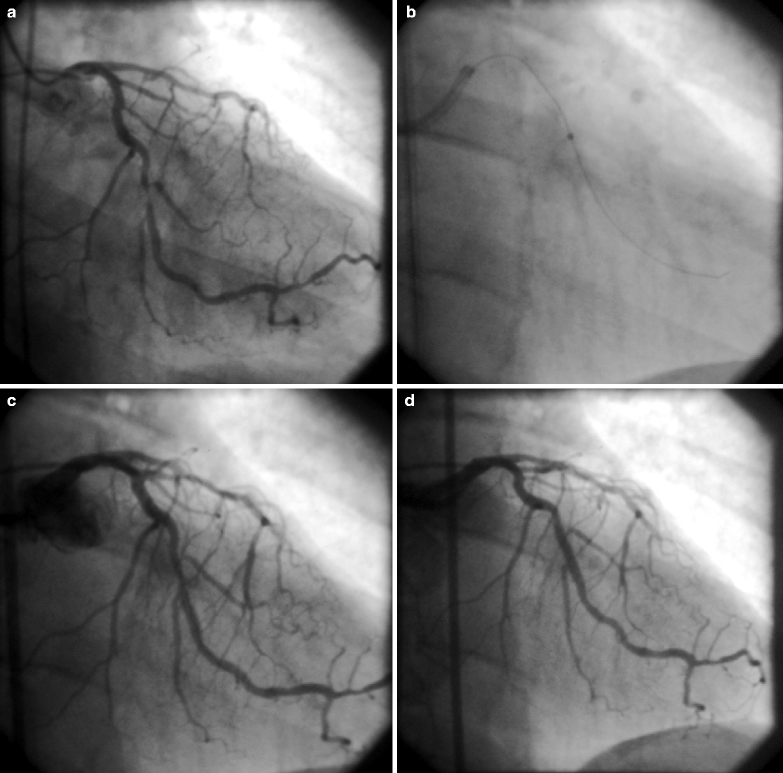
Table 1 presents the current indications for use of laser in cardiovascular medicine. Careful case selection is integral to ensuring successful laser procedures. In acute ischemic coronary syndrome caused by obstructive plaque and associated thrombus, restoration of normal antegrade coronary flow is crucial for preservation of the myocardium. As the laser devices interact uniquely with plaque and thrombus, they can be successfully applied in select patients who sustain unstable angina pectoris (Fig. 1) or acute myocardial infarction.13 The target vessel in these clinical scenarios is either a native coronary artery or an old saphenous vein graft. An important advantage of the laser is its safe activation in patients with depressed left ventricular ejection fraction presenting with acute ischemic coronary syndromes. Both holmium:YAG14 and excimer laser have been demonstrated to be successful revascularization modalities for compromised ventricular function. When the outcome of excimer laser assisted angioplasty and stenting was compared in patients with depressed ejection fraction (mean LVEF = 28 ± 6%) versus those with preserved ejection fraction (LVEF = 53 ± 8%), a successful debulking and thrombus removal occurred regardless of baseline ventricular function.15

Table 1
Current indications for use of laser in cardiovascular medicine
1. Acute coronary and peripheral ischemic syndromes |
2. Symptomatic patients with evidence of coronary and peripheral atherosclerotic disease |
3. Treatment of complex coronary and peripheral atherosclerotic lesions – thrombotic, concentric, eccentric, sub-total and total occlusions |
4. Revascularization of native coronary and peripheral arteries and old saphenous vein grafts |
5. Extraction of pacemaker and defibrillator leads |
6. TMLR for treatment of refractory, debilitating angina |

Fig. 1
Application of excimer laser in a patient with unstable angina pectoris. (a) Angiogram demonstrating a 95% eccentric, thrombotic stenosis at the middle portion of a large obtuse marginal branch of the Circumflex artery. (b) A 1.4 mm COS excimer laser (Spectranetics, Colorado Springs, CO) at the target lesion. (c) Results post successful antegrade and retrograde laser debulking along the stenosis. (d) Final angiogram following adjunct stenting with a drug eluting stent. There is complete patency of the target vessel. The patient recovered clinically
Contraindications
Lack of informed consent, unavailability of surgical coverage and unprotected left main disease (a relative contraindication) are considered coronary contraindications. As for peripheral laser applications, the presence of poor flow in a sole remaining vessel to the lower limb constitutes a contraindication.
The Specific Effects of Laser on Thrombus
Thrombus is commonly found in patients with unstable angina or acute myocardial infarction. The presence of intracoronary thrombus is associated with an increased complication rate both during and after percutaneous interventions. Laser energy interacts with two essential components of thrombus: fibrin and platelets. Pulsed wave lasers such as the mid infrared holmium:YAG and the ultraviolet excimer create acoustic shock waves that propagate along the irradiated vessel. These waves carry a dynamic pressure front toward the fibrin mesh within the thrombus. This process disrupts and breaks the fibrin fibers resulting in fibrinolysis and thereby reduces thrombus size.16 Clinically, the excimer laser has been found to be a useful interventional tool for targeted thrombus removal strategy.17 This laser also alters the aggregation kinetics of platelets leading to reduced platelet force development and inhibition of platelet activity. This phenomenon of platelets stunning is dose dependent and most pronounced at high fluence levels such as 60 mJ/mm2.18
Laser in Acute Myocardial Infarction
Patients who sustain acute myocardial infarction and continue to experience persistent chest pain frequently exhibit an unstable hemodynamic condition. These patients commonly present after a failed response to thrombolytic pharmacotherapy or have contraindications to these medications. In such circumstances, a rescue intervention is indicated for preservation of myocardial tissue. The interest in excimer laser as a potentially beneficial revascularization modality in these patients stems from recognition of its potential for thrombus removal and concomitant plaque debulking. The CARMEL (Cohort of Acute Revascularization of Myocardial infarction with Excimer Laser) multicenter study enrolled 151 patients with acute myocardial infarction and large thrombus burden was present in the infarct related vessel in 75% of the patients. The excimer laser restored an abnormally low baseline coronary TIMI flow in the infarct related vessel (native coronary artery or a saphenous vein graft) to a normal level (Grade 3 TIMI flow) of antegrade perfusion. It also successfully debulked and decreased the target stenosis which was followed by adjunct stenting. An overall 91% procedural success rate, 95% device success rate and 97% angiographic success rate were reported.19 A low rate (8.6%) of major adverse coronary events was recorded. Specifically, 3% dissection and only 0.6% distal embolization rates were encountered. There were no laser induced perforations. Death occurred only among 30% of the patients presenting with cardiogenic shock. Importantly, maximal laser effect was observed in lesions laden with a heavy thrombus burden. Separate analysis of the study’s data base demonstrated maximal laser gain among those patients who presented with an already established Q wave myocardial infarction, an ongoing ST segment elevation and large-extensive thrombus burden in the infarct related vessel.20 Altogether, the excimer laser is capable of removing as much as 80% of the thrombus burden from the treated targets.21
Application of Laser in Specific Target Lesions
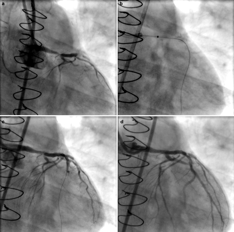
Left main disease: Until recently left main coronary artery disease was considered a contraindication for percutaneous intervention. However with a recent paradigm shift from surgical approach to percutaneous revascularization, a defined role for laser in such a strategy has emerged. In a series of 20 symptomatic patients with severe left main disease, the excimer laser was used for pre-stent debulking (Fig. 2). Traditionally, performing a safe percutaneous intervention in the left main coronary artery requires at least partial myocardial protection by patent bypass grafts. However, in this experience patent grafts were present in only 20% of these patients while the majority had no protection or poor protection by a diseased graft. Nevertheless, successful intervention was achieved in 95% of the patients. The investigators concluded that small size laser catheters, when used with proper lasing technique and adjunct stenting can lead to successful debulking strategy in select patients with left main stenosis.22


Fig. 2
Application of excimer laser in left main coronary artery disease in a patient with unstable angina. The patient underwent coronary bypass surgery just 4 weeks earlier but two saphenous vein grafts occluded with only the left internal mammary artery remaining patent. (a) The critical stenosis at the distal segment of the left main coronary artery. (b) Activation of a 0.9 mm X-80 excimer laser (Spectranetics, Colorado Springs, CO) catheter (the tip of the catheter is radiologically opaque) along the plaque. (c) Adequate recanalization post laser debulking. (d) Final angiographic results post adjunct stenting: patent left main artery is present with adequate flow into the Circumflex artery. The patient’s symptoms were completely relieved
Undilatable or uncrossable lesions: Excimer laser can be very useful in the treatment of atherosclerotic lesions which cannot be crossed with conventional balloon systems. It can also be effectively applied in lesions that fail to yield to the displacement forces induced by balloon angioplasty. The success rate in uncrossable or undilatable stenoses is high, approaching 90%. However, when these targets are calcified, the response is less favorable to laser debulking than that of non calcified lesions (79% vs. 96%, p < 0.05).23
Calcified lesions: Balloon angioplasty frequently produces suboptimal results in these lesions. In the past, lasers utilizing a wide range of wavelengths encountered difficulties in recanalization of these stenoses. However, following Rentrop’s invention of a high energy level excimer laser catheter (capable of producing up to 80 mJ/mm2 at 80 Hz energy fluence) for calcified lesions, the device was introduced to the field as the 0.9 mm X-80 catheter. Improved results have been reported with this technology.24 The unique capability of the excimer laser to debulk underlying calcium is also demonstrated in the critical scenario of a non-dilatable coronary stent. In such challenging cases calcium impinges on the stent struts and obstructs their full expansion during deployment. Laser ablation either softens or removes the calcium, thus enabling subsequent complete deployment and proper expansion of a stent.25
Total occlusions: These challenging atherosclerotic stenoses are frequently difficult to traverse with a guide wire, respond unfavorably to balloon angioplasty and resist stent deployment. A laser catheter or a laser wire can penetrate these lesions and facilitate adjunct balloon and stenting.26 A success rate of 86–90% for the excimer laser has been reported in these occlusions.27 Total occlusions are mostly long standing lesions that contain layers of old, well organized thrombus within calcified and fibrotic plaques. In the setting of acute myocardial infarction, however, they can present a specific revascularization challenge because of a large, fresh thrombus burden as imposed on an underlying plaque material. In such instances the laser can be used successfully for recanalization of the total occlusion.28
Aorto-ostial lesions: These resistant atherosclerotic obstructions are usually focal and often calcified. Precise laser debulking enables subsequent stenting. The success rate with laser and adjunct balloon angioplasty in two series of patients were reported as high as 94%,29,30 thus, far exceeding the relatively low 74–80% success rate for standard balloon angioplasty.
Saphenous vein grafts lesions: Occlusions in old saphenous vein grafts frequently consist of diffuse or multifocal plaques and thrombus. These lesions are degenerative and prone to distal embolization. Despite the presence of a large thrombus burden within these vessels, a success rate of 94% was reported with both the ultraviolet excimer laser and mid-infrared holmium:YAG laser.31,32 The considerable capability of laser to provide safe debulking of these grafts even in the setting of acute myocardial infarction and in the presence of a heavy thrombus burden has been documented.33 Furthermore, the remarkably low rate of distal embolization during excimer laser debulking of old bypass grafts (1–5%) precludes the need for adjunct protection systems in most cases where the excimer laser is used.
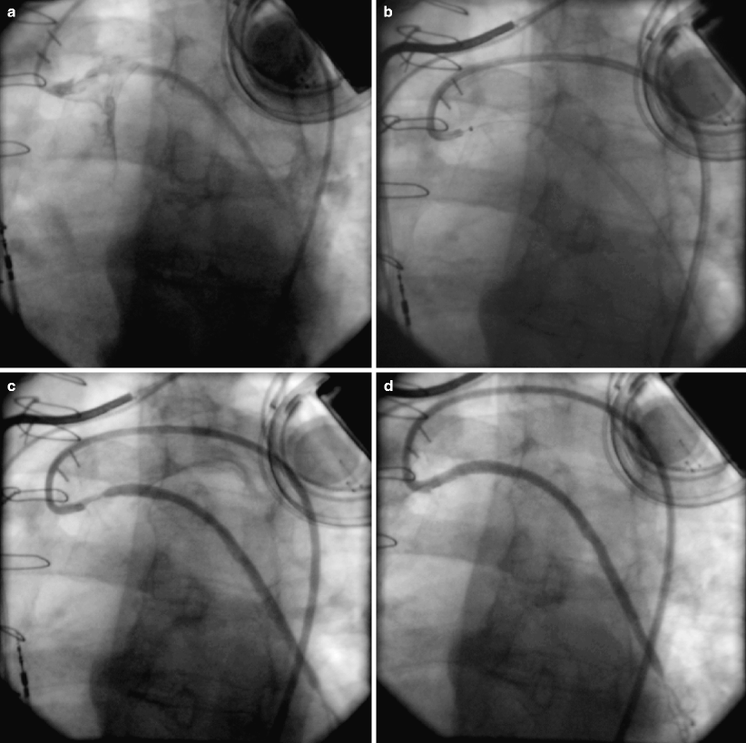
Stent restenosis: Stent restenosis is attributed to localized or diffuse tissue growth. Laser debulking of stent restenosis is a preferred treatment over tissue displacement by standard balloon angioplasty.34 Mehran et al. compared excimer laser and adjunct balloon dilations to balloon treatment alone and concluded that no complications were associated with the use of laser. The laser application resulted in greater luminal gain, ablation of intimal hyperplasia, and a tendency toward less frequent subsequent target vessel revascularization.35 In cases of focal or severe diffuse stent restenosis within old vein grafts, laser debulking and removal of the obstructive regrowth tissue and its associated thrombus facilitates adjunct balloon angioplasty (Fig. 3).


Fig. 3
Application of excimer laser in a critically ill patient with severe angina, marked anterior –lateral ischemia, unstable hemodynamic condition, impaired renal function and depressed left ventricular function. Initial angiography demonstrated critical disease in two old saphenous vein grafts to the obtuse marginal branch of the Circumflex artery and to the left anterior descending artery, respectively. A third bypass graft to the right coronary artery was totally occluded for several years. Of note, this patient underwent coronary bypass surgery twice in the past. A third open heart surgery was not considered a treatment option. Revascularization of the graft to the obtuse marginal artery: (a) A 99% stenosis of the proximal graft. (b) 0.9 mm X-80 excimer laser catheter at the stenosis. (c) Creation of a “pilot channel” along the target lesion. (d) Adequate results following adjunct stenting with a drug eluting stent
Trans Myocardial Laser Revascularization (TMLR)
This unique revascularization concept is based on the diversion of arterial blood flow onto regions of the myocardium which do not receive adequate perfusion secondary to atherosclerotic coronary arterial disease. The attempts to reperfuse ischemic myocardium by direct myocardial recanalization predate coronary bypass grafting and coronary balloon angioplasty.36 The TMLR procedure aims to create multiple 1 mm intramyocardial laser channels (TMLR) within the ischemic viable myocardium. The creation of these channels is expected to promote angiogenesis with growth of microvessels that provide fresh blood supply to the myocardium.37 The increase in microvessels significantly correlates with the expression of matrix metalloproteinases-2 and platelet-derived endothelial cell growth factor.38 A specific indication of the TMLR is for patients with refractory angina pectoris who are unable to undergo coronary bypass surgery for pain relief. TMLR has also been incorporated as an adjunct surgical treatment for those who undergo coronary bypass surgery but need further intraoperative revascularization of myocardial regions that cannot be reached with bypass grafts. The TMLR application involves application of specially designed laser catheters under direct vision or under fluoroscopy guidance. These catheters are placed in direct contact against the epicardium and activated with emission of laser fluence inward toward the myocardium. Various laser sources such as CO2, excimer and holmium:YAG have been used for TMLR either surgically or percutaneously for treatment of patients with refractory angina.39 TMLR has been shown in randomized studies to improve subjective symptoms of40,41 promote increased exercise time42 and improve quality of life as compared with maximal medical therapy.43 Recently it has been proposed that combining TMLR with cell therapy delivered through direct myocardial injections is a safe strategy which may act synergistically to reduce myocardial ischemia and improve functional capacity.44 However, despite a certain level of success TMLR remains quite an enigma as the exact functional mechanism of angina relief is unclear and the relief of angina may be short lived in many.45 The prevailing theory of laser induced thermal damage to cardiac nerves resulting in cardiac denervation has been refuted.46 Furthermore, whether TMLR results in improved cardiac function remains a controversial issue.
Laser for Revascularization of Heart Transplant Allograft Vasculopathy
Stay updated, free articles. Join our Telegram channel

Full access? Get Clinical Tree


