Impaction Bone Grafting of the Acetabulum

CHAPTER 49 Impaction Bone Grafting of the Acetabulum


Total hip arthroplasty (THA) is one of the most successful procedures in modern medicine, and the number of patients receiving a total hip implant is increasing every year. However, this also indicates that the number of patients who need a revision of a previously implanted total hip is increasing. In the long term, the main reason for failure of all types of total hip implants is aseptic loosening. Other reasons for failure are septic loosening, recurrent dislocation, malposition, periprosthetic fractures, and mechanical failure of the implant. In most cases failure leads to bone stock loss, and revision surgery in cases with extensive bone stock loss is demanding. In general the outcome of a revision of a failed hip implant is less successful in those hips with the greatest bone stock loss.


On the acetabular side, the loosening process can result in a cavitary bone defect, but in the more serious cases segmental wall defects also develop in combination with a cavitary bone deficiency. Many acetabular reconstruction techniques have been described both with cemented and noncemented cups. The best approach to these bone stock deficiencies is still under debate and depends not only on the quality of the remaining bone and the extension of the bone loss, but also on the experience of the surgeon with a certain technique.


In Nijmegen we have a long history of reconstructing deficient acetabula with the impaction bone grafting (IBG) technique, both in complex primary and revision hip surgery. We use tightly impacted morcelized cancellous autograft and allografts in combination with a cemented cup in all cases in which acetabular bone stock loss is present. We believe that this is an attractive biologic technique because with this method one really can restore the damaged bone stock. If further future revisions become necessary, which can be expected because in time all arthroplasties will fail, the situation of the bone stock is improved and a second revision becomes easier to perform.


We have used the IBG technique to reconstruct acetabular bony defects since 1979 and frequently have reported on the clinical outcome in patients with primary THA,1 in young patients,2 in patients with congenital dysplasia of the hip (CDH),3 in revision THA,4 and in patients with rheumatoid arthritis.5 In general the clinical outcomes are favorable, even with long-term follow-up. IBG is not an easy and straightforward technique, and success depends on performance of the technique used by the surgeon. The surgeon should understand the basic principles of the technique. An inferior technique will lead to early instability and migration of the acetabular components We believe that a thorough understanding of the technique of containing and impacting morcelized bone grafts will optimize the clinical outcome for the patients.


IBG is a biologic method that can reconstruct bone stock loss. Solid impaction with a hammer and impactors is mandatory. Wire meshes are needed to reconstruct segmental bone defects. When considering IBG, starting with simple cavitary defects to become familiar with the technique and instruments is recommended.


Optimal cementation techniques and a proven implant are mandatory.


The purpose of this chapter is to discuss the most important mechanical and technical aspects of acetabular IBG and to provide the guidelines for an adequate surgical technique to reconstruct a deficient acetabulum in combination with a cemented component.


INDICATIONS AND CONTRAINDICATIONS


IBG is used in our institution in all patients with acetabular bone stock loss who are scheduled for total hip reconstruction, both in difficult primary cases and revision cases.


In primary total hip reconstruction with simple protrusio acetabuli, we use the autogenous femoral head as the source of bone chips, sometimes in combination with trabecular bone from the proximal femur. However, in primary cases with severe bone stock deficiencies (e.g., CDH) we also use fresh frozen femoral head allografts in combination with the patient’s own femoral head and wire meshes to construct a new acetabular wall at the anatomic center of rotation.


In revision THA with bone stock loss, it is essential to detect the reason for failure. The treatment of septic loosening is essentially different from that of aseptic loosening. Septic loosening is one of the main contraindications for the technique. Before reconstruction with the IBG technique is considered, every effort should be made to exclude infection. In cases in which septic loosening is suspected, laboratory tests, technetium scanning, gamma-immunoglobulin scintigraphy, and preoperative aspiration of the hip to obtain material for culturing should be used to detect if infection is the reason of the loosening. In septic loosening, the existing infection should first be treated by surgical means and medication. IBG can be used in cases of sepsis but only after proper treatment of the infection with antibiotics using a two-stage procedure.


There are many alternative options for acetabular revisions. Cement-only reconstructions may be indicated in the elderly with a limited life expectancy. IBG may be not needed, and the surgical procedure is too extensive for the elderly with a shorter life expectancy. Many different techniques and implants for noncemented reconstructions are also available. Impacted bone grafting has also been described in combination with metal shells, solid reconstruction rings, and noncemented cups. We have no experience with these methods. Seemingly small modifications of the original technique of IBG using the wire meshes and a cemented cup are not recommended. Although such modifications have been tried, the reported outcome has not always been favorable.


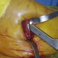
In acetabular revisions with pelvic dissociation it is mandatory to stabilize this pelvic fracture first. Reconstruction of such a dissociation using only flexible wire meshes will fail. Flexible wire meshes do not result in not proper osteosynthesis. The meshes are too thin and flexible, and fixation using small fragment screws is inadequate for the stabilization of the pelvic fracture. Proper pelvic plates and screws must be used to fix and stabilize adequately. Only after the fracture has been fixed can wire meshes be applied to cover the segmental defects and create a cavitary defect that can be filled and impacted with morselized bone chips (Fig. 49-1).


Bone impaction grafting will fail in a high percentage of patients with bone stock loss or failed hips resulting from pelvic radiation therapy. Dead pelvic bone is not a suitable host bone bed for cancellous bone ingrowth, and also the infection rate is unacceptably high.


Basic knowledge of acetabular cementation techniques is mandatory when IBG is used; otherwise a good result cannot be expected.


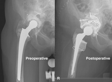
PREOPERATIVE PLANNING


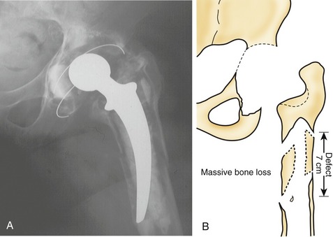
In the planning of revisions it should be realized that bone stock defects and acetabular distortions encountered during surgery are frequently much more severe than the preoperative radiographs suggest. Good-quality plain radiographs in two directions are therefore necessary to evaluate the severity of the anatomic distortion, the location and extent of bone lyses, and, if appropriate, the bone cement distribution. A good rule of thumb is that plain radiographs show only 50% of the reality (Fig. 49-2).


If plain radiographs in two directions are not sufficient to establish the extent of the deficiencies, a CT scan can be considered.


Preplanning of the proposed implant can best be done using the contralateral side. Especially if the contralateral hip is not affected or if a well-performing total hip implant is in situ, templating on the contralateral side is valuable. In this way equal leg length and the creation of the appropriate offset are best achieved. Templating on the affected side will provide more insight regarding the bone loss, prepares the surgeon and surgical team for the extension of the surgery, and gives a good estimate of materials needed: wire meshes, plates, screws, and amount of bone graft.


Important, as mentioned before, are the patient’s age, his or her physical condition, and the existence of associated diseases. Also, results of laboratory results should be taken into consideration (e.g., erythrocyte sedimentation rate, white blood cell count, C-reactive protein, hemoglobin, hematocrit). If the patient is not fit to undergo extensive surgery or the life expectancy is too low, IBG is not the appropriate treatment.


Mar 10, 2016 | Posted by in Reconstructive surgery | Comments Off on Impaction Bone Grafting of the Acetabulum

Full access? Get Clinical Tree

Get Clinical Tree app for offline access