Fig. 3.1
Progression of infantile hemangioma on the right side of the face at 1 month in the first visit (left) and at 1.5 years (right). The ulcers in the preauricular, the cheek, and the submandibular areas are healed
Vascular malformations consist of capillary malformation (CM), venous malformation (VM), lymphatic malformation (LM), and arteriovenous malformation (AVM). Some clinically combine more than one malformation and then categorize complex vascular malformation and complex syndromes such as Klippel-Trenaunay syndrome (CM + VM + LM) or Parkes Weber syndrome (AVM/or arteriovenous fistula (AVF) + skin pseudo-CM + lymphedema) as observed in more systemic signs and symptoms.
Skin necrosis often manifested in severe AVM and combined CM + LM as well as minority cases of IH.
3.2 Assessment and Imaging Tools
Many imaging tools are able to determine the diagnosis of vascular malformations.
3.2.1 Conventional X-Rays
These are usually of no or little value in most cases. Venous malformations (VMs) may be diagnosed if phleboliths (vascular stones) are observed on plain X-rays. Bone distortion is merely seen in large malformations with a soft tissue mass effect. Some diffuse and massive VMs cause osteolytic lesions and bring about a risk of pathologic fractures. AVMs involving the bone sometimes lead to osteolytic lesions due to intraosseous nidus or large draining venous channels after nidus.
3.2.2 Duplex Ultrasonography
This imaging is primarily used as a diagnostic tool [5] at the clinic in the first visit. It permits distinction between tumors and malformations. It also identifies a vascular malformation and pinpoints the type of lesion. It demonstrates either the lesion is cystic or tissular, clarifies either the presence or absence of flow, and thus distinguishes between fast-flow and slow-flow malformations (Fig. 3.2). Angiostructure and vessel density can be assessed; however, its reliability is most of the time poor. Peak flow velocities and arterial output may be measured in AVMs. In head and neck or extremity AVMs, comparing arterial output on the normal side and the abnormal contralateral side is crucial in prognosis, especially of possible cardiac failure, and thus useful for follow-up of AVMs.
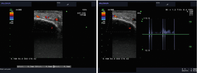
Fig. 3.2
Typical duplex ultrasonic examination. Flow patterns and localization of the lesions are depicted (left); flow intensity and pulse are demonstrated (right)
3.2.3 Computed Tomography (CT)
This method is relatively of limited interest even with enhanced contrast, information of a lesion is merely whether highly vascularized or not. Precise delineation and diagnosis of soft tissue lesions remain frail with the exception of macrocystic LMs, where cysts are clearly depicted. The presence of phleboliths may lead to a diagnosis of venous malformation as distinctive calcifications develop on thrombosis and debris resulting from slow flow. Bony displacement or alteration can be seen due to chronic (long-term) compression seen in both VMs and LMs. Pathologic fractures and absorption may be seen in bone or bone-adjacent AVMs.
3.2.4 Magnetic Resonance Imaging (MRI)
This is the best diagnostic modality, giving optimal analysis of soft tissue masses and proper diagnosis, distinguishing tissular form cyst, and delineating fast or slow vessel flows. Venous and lymphatic malformations have each attribute pattern. They are hyperintense on spin echo T2-weighted sequences and optimally seen in fat-suppression sequences. T1-weighed and fat-suppression sequences with gadolinium injection demonstrate an intense enhancement in infantile hemangioma, while the enhancement is inconsistent and progressive on dynamic sequences in VMs. Gadolinium contrast allows differential diagnosis among VMs and LMs. LMs can be distinct from VMs as LMs indicate enhancement only at the margins of the cysts. By contrast, VMs are clearly and evenly stained. MRI is compulsory before treatment to make decisions of the extent of the lesion and the relationship among the vascular malformation, intact neighboring nerves, and vessels and for the identification and diagnosis of the lesions. In fast-flow vessels, they are identified as flow voids. MR angiography can confirm the diagnosis of fast-flow pathology; however, it remains insufficient for detecting accurate AVM’s nidus and angiostructures.
Related posts:


Stay updated, free articles. Join our Telegram channel

Full access? Get Clinical Tree


