Time of onset of clinical signs of skin injury depends on the dose received.
10.3 Radiation Ulcers
Radiation treatment causes endothelial damage and fibrosis, leading to impairment of vascular and lymphatic flow. This impairment produces hypoxic, hypocellular, and hypovascular tissue, which is unable to maintain normal tissue turnover, resulting in tissue necrosis, infection, and ulceration [5].
Dry and moist desquamations are skin clinical manifestations of the keratinocyte mitotic death, and necrosis is the consequence of manifestations of tissue subcutaneous structures such as the muscles, vessels, and sometimes even bone [6].
10.4 Management of Radiation Ulcers
10.4.1 Debridement
Local wound care with standard wet or wet-to-dry dressings is rarely successful in promoting satisfactory debridement, cleansing of the wound, and subsequent growth of supported granulation tissue.
Radiation ulcers frequently progress and become much worse over time. The underlying ischemia sets in motion a cycle of infection and necrosis, leading to the development of additional ulcers in the surrounding tissue and finally to frank gangrene. Debridement of all nonvital tissue is required at the first stage of treatment. When the damage extends deep into the muscles or bones, extensive debridement should be done [7].
10.4.2 Methods of Wound Closure
10.4.2.1 Surgical Treatment
Radiation ulcers will not heal by aggressive medical wound management. Skin grafts and local cutaneous flaps located within the radiation field are unreliable and rarely provide adequate stable coverage because of the poor state of the wound bed. Salvage surgery with musculocutaneous flap is the recommended method to heal these complex wounds.
Case 1
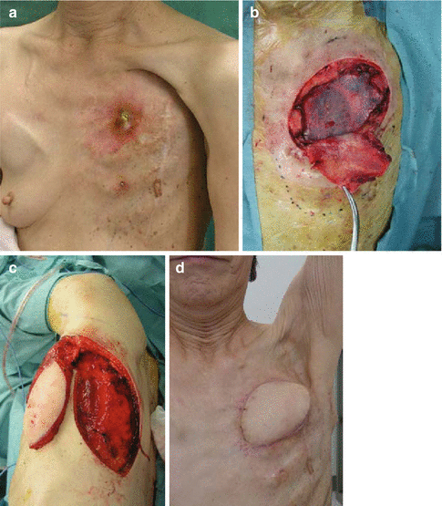
A 77-year-old female underwent an unknown quantity of radiation treatment due to breast cancer after ablation of the tumor. Chronic chest ulcers, which formed fistulae penetrating to the rib, were observed. Intraoperative examination of surgery showed necrotic ribs, which were ablated with the parietal pleura. The pleura was restored using artificial substances. The radiation ulcer was reconstructed without disturbance of shoulder movement.
Necrotizing rib was exposed (Fig. 10.2a). After debridement, the pleura was restored using artificial substances (Fig. 10.2b). Latissimus dorsi musclo-cutaneous flap transfer from the back (Fig. 10.2c). Six months after the surgery, the wound was resurfaced (Fig. 10.2d)


Fig. 10.2


A 77-year-old female underwent an unknown quantity of radiation treatment due to breast cancer after ablation of the tumor. Necrotizing rib was exposed (Fig. 10.2a). After debridement, the pleura was restored using artificial substances (Fig. 10.2b). Latissimus dorsi musclo-cutaneous flap transfer from the back (Fig. 10.2c). Six months after the surgery, the wound was resurfaced (Fig. 10.2d)
Stay updated, free articles. Join our Telegram channel

Full access? Get Clinical Tree


