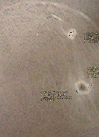
Fig. 38.1
(a, b) Microcalcifications located at 1 o’clock mediocranial in the right breast

Fig. 38.2
Breast MRI showing at least two distinct lesions
38.2 Surgery
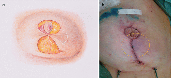
After preoperative drawings were done, the nipple-areola complex was supplied by a lateral pedicle (modified Hall Findlay). After resection of the tumor, the skin was closed again, however, with strong tension on the edges (Fig. 38.3a). After closure of the vertical scar, the tension resulted in a hypoperfused area along the scar (Fig. 38.3b). We decided to use the vertical reduction technique as the patient had no ptosis and with medium- to large-sized breast with a jugulo-nipple distance of 28 cm and a submamarian-nipple distance of about 9 cm. Our primary intention was to remove the mediocranial quadrant and fill the defect with the central and lower portion of the breast. Moreover with the Hall Findlay technique, scars are avoided at the medial and inner quadrant (no man’s land).
Get Clinical Tree app for offline access


