Chapter 59 Geriatric dermatology
1. How common are skin disorders in the elderly population?
Survey studies have demonstrated that skin diseases are more common in the geriatric population than in the general population. One study revealed that 40% of Americans between the ages of 65 and 74 years had a cutaneous disease significant enough to warrant treatment by a physician. Patients older than 74 years are even more likely to develop significant skin diseases.
2. What is intrinsic aging of the skin?
Aging of the skin may be divided into that due to intrinsic aging and that secondary to extrinsic aging (Table 59-1). Intrinsic aging includes those changes that are due to normal maturity and senescence and thus occurs in all individuals. Classically, intrinsic aging has not been considered to be preventable, but there is renewed interest in the role of antioxidants, such as vitamins C and E, in preventing intrinsic aging. Despite numerous articles in the lay literature, there is no proof that these treatments are effective.
3. What is extrinsic aging of the skin?
Extrinsic aging of the skin consists of those changes produced by external agents. The most important extrinsic factor is cumulative ultraviolet (UV) light exposure. The cutaneous changes produced by sunlight are collectively referred to as dermatoheliosis. Most of the changes associated with aging of the skin, such as wrinkles, yellow leathery skin, thin skin, hyperpigmentation, hypopigmentation, lentigo senilis (liver spots), telangiectasias, and senile (solar) purpura, are all secondary to damage from the sun or other UV light sources such as tanning booths. Less important extrinsic agents that accelerate aging of the skin include smoking and possibly environmental pollutants.
Table 59-1. Age-Related Changes in the Skin
| INTRINSIC AGING | EXTRINSIC AGING (PRIMARILY UV LIGHT) |
|---|---|
4. How does intrinsically aging human skin vary from young skin under the microscope?
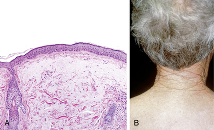
Microscopically, the epidermis in aged skin demonstrates flattening of the dermoepidermal junction with loss of the normal rete ridge pattern (see Fig. 59-2A) with fewer melanocytes and Langerhans cells. The dermis demonstrates atrophy with fewer fibroblasts, mast cells, and blood vessels associated with depigmentation of hair, loss of hair follicles, and fewer sweat glands. The amount of collagen, elastin, and ground substance also decreases.
5. Why does skin wrinkle as we age?
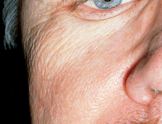
The answer is complicated, because some authorities recognize as many as five different subtypes of wrinkles. The most common type of wrinkles on non–sun-exposed skin are fine wrinkles (glyphic wrinkles) that represent accentuation of normal skin markings. Microscopically, this is due to focal thinning and decreased numbers of keratinocytes. This appears to be intrinsic to aging (Fig. 59-1). The deeper wrinkles in photodamaged skin demonstrate a groove in the epidermis associated with solar elastosis that protrudes on both sides of the groove. These deeper wrinkles are due to extrinsic aging, primarily resulting from ultraviolet light.
6. Does smoking cigarettes accelerate skin aging?
Yes. Epidemiologic studies clearly indicate that smoking is an important extrinsic factor in accelerating aging of the skin. This is also support by animal studies and in vitro studies that have demonstrated that smoking increases the production of tropoelastin and matrix metalloproteinases, resulting in increased degradation of matrix proteins (e.g., collagen, elastic fibers) and abnormal production of abnormal elastotic material. Just another reason not to smoke!
Morita A: Tobacco smoke causes premature skin aging, J Dermatol Sci 48:169–175, 2007.
7. What is solar elastosis?
Solar (actinic) elastosis refers to the changes due to abnormal elastotic fibers (Fig. 59-2A) produced by fibroblasts in the papillary and superficial reticular dermis in response to UV light exposure. These abnormal elastotic fibers stain with elastic tissue stains; electron microscopy demonstrates that these fibers are similar, but not identical, to normal elastic fibers. Recent research suggests that they are the result of UVA damage to fibroblasts that results in the over-production and accumulation of elafin, which binds to elastic fibers making them resistant to normal degradation by elastase. Large aggregates of these fibers impart a yellowish color and account for the yellow leathery appearance of sun-exposed skin in geriatric individuals. Solar elastosis is often most easily appreciated in the posterior neck, where it is termed cutis rhomboidalis nuchae (Fig. 59-2B).
8. What is nodular elastosis with cysts and comedones?
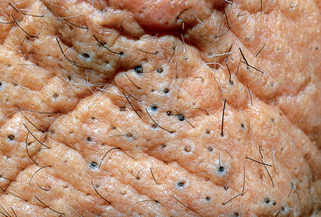
Nodular elastosis with cysts and comedones, also known as Favre-Racouchot syndrome, is characterized by the presence of marked solar elastosis and comedones on the lateral and inferior periorbital areas (Fig. 59-3). Severe cases may demonstrate cysts. The reason for this regional presentation is not understood, but it has been suggested that the fibroblasts around the hair follicles are damaged by UV light and no longer produce normal elastic tissue. This predisposes to dilatation of the hair follicles, resulting in comedones and cysts. Most cases can be successfully treated with topical tretinoin cream and comedonal extraction.


Stay updated, free articles. Join our Telegram channel

Full access? Get Clinical Tree


