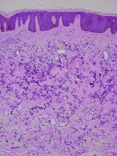
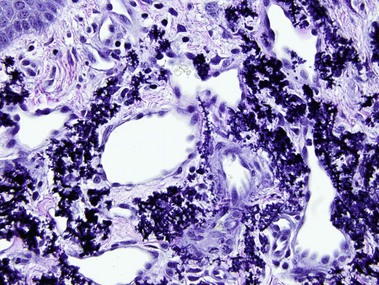
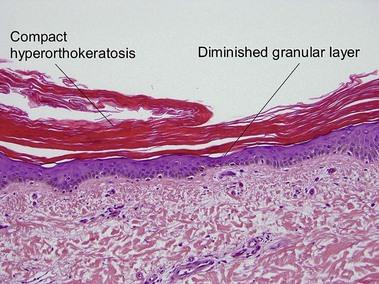
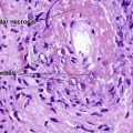
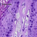
Genodermatoses

Chapter 12


Genodermatoses


image


A summary table of the ‘Key histologic features of genodermatoses’ can be found in the on-line content for this book.


Pseudoxanthoma elasticum


Pseudoxanthoma elasticum is an autosomal-recessive and, less commonly, autosomal-dominant disorder due to a mutation in the ABCC6 transporter gene. It results in calcification of the elastic fibers of the skin, eyes, and artery walls. The skin changes become apparent around the second decade and simulate “plucked chicken skin” in the flexural areas. Biopsy of a scar may reveal the elastic tissue abnormalities in a patient with no cutaneous lesions. Eye changes include angioid streaks and retinal hemorrhage that may cause blindness. The vascular changes may lead to hypertension, stroke, myocardial infarction, mitral valve prolapse, and gastrointestinal hemorrhage.


Similar histologic findings can also be seen after topical exposure to calcium salts. Periumbilical perforating pseudoxanthoma elasticum (perforating calcific elastosis) affects multiparous black women but is limited to an isolated area. Patients on long-term penicillamine for Wilson’s disease may develop altered elastic fibers with overlapping features of pseudoxanthoma elasticum and elastosis perforans serpiginosa. The penicillamine-induced, altered elastic fibers have small lateral buds arranged perpendicularly to the primary elastic fiber resembling the twigs on a bramble bush.


Ichthyosis vulgaris


In most other conditions, hyperorthokeratosis is associated with a prominent granular layer and parakeratosis is associated with a diminished or absent granular layer. Ichthyosis vulgaris (in which there is little to no granular layer despite hyperkeratosis) and axillary granular parakeratosis (in which there is a granular layer despite the presence of parakeratosis) are exceptions to this rule.


Ichthyosis vulgaris is an autosomal-dominant disorder with retention of the stratum corneum (rather than hyperproliferation) resulting in hyperkeratosis. There is a deficiency in profilaggrin resulting in inadequate keratohyaline granule synthesis. There are fine brown to transparent scales that spare the flexures. Accentuation of the palmoplantar creases, keratosis pilaris, and coexisting atopic dermatitis may be present.


Acquired ichthyosis in association with malignancy, especially Hodgkin’s lymphoma, may clinically and histologically mimic ichthyosis vulgaris. Acquired ichthyosis may also occur with niacin therapy.


Incontinentia pigmenti (Bloch–Sulzberger syndrome)


Apr 26, 2016 | Posted by in Dermatology | Comments Off on Genodermatoses

Full access? Get Clinical Tree

Get Clinical Tree app for offline access