(1)
Department of Dermatology, Drexel University, Philadelphia, Pennsylvania, USA
Abstract
The majority of patients with eczema have a filaggrin defect, most commonly mutations R501X and 2282del4, both of which relate to skin barrier function. The truest expression of filaggrin deficiency is ichthyosis vulgaris, and patients who have both ichthyosis vulgaris and eczema have profound itching. Patients with other types of ichthyosis may also have eczema though the causative genes are dissimilar. The common link in these differing genes is their role in making a faulty stratum corneum, preventing it from serving as a barrier to protect the human organism from outside substances. The hypothesis is that biofilm-producing staphylococci that are normal flora under the influence of sweating and occlusion produce biofilms. These biofilms obstruct the sweat ducts and lead to the activation of Toll-like receptor 2, which triggers the production of many mediators, including tumor necrosis factor α, which leads to epidermal spongiosis, and protease-activating receptor 2 (PAR2), which in turn leads to itching. Therefore in this “double-hit” (genetic and environmental) phenomenon, the environmental hit is staphylococci and their biofilms and the genetic hit is filaggrin.
Keywords
aap geneArylsulfatase CDouble-hit phenomenon FALDH (fatty aldehyde dehydrogenase)FilaggrinGenetic “hit” GJB2 gene icaD geneIchthyosis vulgarisKID syndrome (keratitis-ichthyosis-deafness)Lamellar ichthyosisNetherton syndromeSjögren-Larsson syndrome SPINK5 (serum proteinase inhibitor Kazal-type 5) Staphylococcus Steroid sulfataseStratum corneumTransglutaminase 1X-linked ichthyosisGenetics is probably the least heretical component of the eczema story. Everyone believes the disease to be genetic, and few doubt that there may be a filaggrin defect in the majority of patients with eczema [1]. Within that filaggrin defect, one interesting fact is that there are multiple substitutions in the DNA of filaggrin associated with eczema [2]. The most common defects are the two prevalent mutations R501X and 2282del4 [2–4]. Both of these relate to skin barrier function, and either may be present in eczema.
It is most interesting that the truest expression of filaggrin deficiency is ichthyosis vulgaris [2] (Fig. 5.1), a disease that involves very distinctive, scaling, dry skin that does not itch. Certain patients with ichthyosis vulgaris have eczema (Fig. 5.2), and those patients itch profusely [5, 6]. Thus, there is a built-in dichotomy in that aspect of the disease.
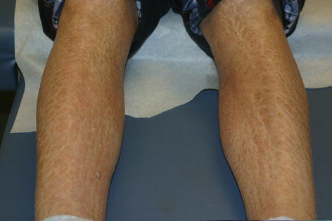
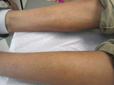
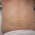
Fig. 5.1
On the legs are plate-like scales; no redness or other sign of inflammation is present. This is the “pure” expression of the filaggrin gene in the disease ichthyosis vulgaris
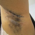
Fig. 5.2
Plate-like scale (a “keyword” for ichthyosis) is present on the bilateral lower extremities. On the right leg are faint pink plaques. This photograph therefore represents both ichthyosis and mild eczema
The other consideration is that patients with different types of ichthyosis such as X-linked and lamellar ichthyosis (Fig. 5.3) may also have associated eczema [7]. These forms of ichthyoses involve defects in genes other than the filaggrin gene, steroid sulfatase for X-linked ichthyosis and transglutaminase1 for lamellar ichthyosis [8]. That these ichthyoses which have differing causative genes can each present with typical forms of eczema is intriguing. It also clearly implies that there is something that each gene does which, although different, allows eczema to develop. In X-linked ichthyosis, the stratum corneum is affected by the accumulation of cholesterol sulfate between the corneocytes, which leads to the clinical findings.
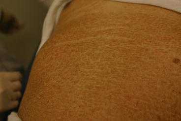
Fig. 5.3
Lamellar ichthyosis. Hyperkeratotic scale is present on the chest in this patient who was born with a collodion membrane
We believe the common link in these differing genes is their role in making a faulty stratum corneum. This prevents the stratum corneum from executing its main function, which is to serve as a barrier to protect the human organism from outside substances [9]. Therefore it is the stratum corneum that “keeps the outside out”; the stratum corneum and the basal zone of the epidermis “keep the inside in.” This is an oversimplification, but it seems a useful concept.
Each of these genes has a somewhat similar role in making the stratum corneum whole. Each also helps make either the material between the corneocytes that binds them together or helps to stabilize the corneocytes themselves. The stratum corneum has consequently been compared to “bricks and mortar,” where the corneocytes are the bricks and the genetically derived material between them is the mortar [10]. Schematically, it appears ceramides are “sandwiched” between hyaluronic acid deposits that line the outer membrane of the corneocytes.
The exact role of the various ichthyosis genes is shown in Table 5.1. Filaggrin binds to and condenses the cytoskeleton of the keratinocytes. It has no clear relationship with ceramide levels in the stratum corneum. Steroid sulfatase, also known as arylsulfatase C, is expressed throughout the body; but, in skin, it breaks down cholesterol sulfate. If this gene is defective, cholesterol sulfate accumulates in the stratum corneum, and there is a scaling skin eruption, a permeable barrier dysfunction, and a retention of corneocytes. Transglutaminase 1 links many proteins together in the corneocyte cell envelope [11]. Each contributes significantly to the “cement” holding these cells together in the stratum corneum [10]. Without them, the skin is excessively dry and scaly.
Table 5.1
Role of 3 ichthyosis genes in stratum corneum formation and associated disease
Gene | Function | Disease |
|---|---|---|
Filaggrin | Filaggrin monomers bind to and condense the cytoskeleton of keratinocytes | Ichthyosis vulgaris |
Atopic dermatitis | ||
Steroid sulfatase (arylsulfatase C) | Breaks down cholesterol sulfate in the epidermis | X-linked ichthyosis |
Transglutaminase 1 | Catalyzes cross-linking of proteins to form a cornified cell envelope | Lamellar ichthyosis |
Related posts:
 Physiology
 Immunology
 Diseases in Which Eczema Is a Secondary Component (Meyerson’s Nevus and Doucas Kapetanakis Pigmented Purpuric Dermatosis)
 The Story of Eczema in Pictures
 Diseases with Occluded Sweat Ducts other than Eczema (Tinea Pedis, Axillary Granular Parakeratosis, and Seborrheic Dermatitis)
 Clinical Presentations
Stay updated, free articles. Join our Telegram channel

Full access? Get Clinical Tree


