Type I
One vascular pedicle, e.g., gastrocnemius muscle, rectus femoris, and tensor fasciae latae
Type II
Dominant vascular pedicle and minor pedicle, e.g., biceps femoris, gracilis, semitendinosus, trapezius, vastus lateralis
Type III
Two dominant pedicles, e.g., gluteus maximus, rectus abdominis, semimembranosus
Type IV
Segmental vascular pedicles, e.g., sartorius
Type V
One dominant vascular pedicle and secondary segmental pedicles, e.g., pectoralis major, latissimus dorsi
The concept of taking vascularized tissue from a distant site for immediate transfer to a pressure ulcer defect with immediate reconnection of the axial artery and vein using microsurgical techniques was described by Chen et al. [12]. This concept, however, is not practical for every pressure ulcer performed on a daily basis and is of limited application.
Transfer of sensory tissue to an insensate area of pressure ulcer in paraplegic patients was attempted by Cochran et al. [13]. Attempts were made by Daniel et al. [14] to reinnervate a flap to restore sensation to the area of pressure ulcer by using a long nerve graft from above the level of the cord injury. These techniques have not been universally successful in achieving and maintaining healing through restoration of sensation.
The introduction of tissue expansion in plastic surgery more than 20 years ago stimulated surgeons to use this technique in closing pressure ulcers. It was described by Esposito [15], who claimed the main advantage was to advance sensory skin to cover an insensate pressure ulcer area. Braddom and Leadbetter [16], Yuan [17], and Neves et al. [18] reported that unstable skin resulted from a healed pressure ulcer or graft or tissue expander are not ideal to cover pressure ulcer used to close a pressure ulcer. The author shares the concern of using a tissue expander as a foreign body near a contaminated wound. In addition, the expanded skin can only cover flat surface wounds and not a cavity, and it requires a long time to achieve expansion of the skin to be utilized for the ulcer closure. From 1980 to the present, there have been hundreds of articles published in plastic surgery journals describing the use of muscle or musculocutaneous flaps in different forms or shapes to close different pressure ulcers in various anatomical parts of the body. Today, the standard surgical method for closing pressure ulcers, which has become the standard of teaching and training for plastic surgery trainees, is to use a muscle or musculocutaneous flap [19–21].
7.3 The Ladder of Reconstructive Surgery

7.4 Principles of Flap Design and Repair of Pressure Ulcers
The specific anatomical details of flap design are different depending on the location of the pressure ulcer, but the principles are the same. The design of the flap in plastic surgery depends on the following considerations:
1.
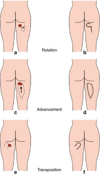
Geometrical design is the way the flap is moved toward the defect by advancement, rotation, or transposition (Fig. 7.1a–f). The flap donor site can be closed directly or, if the donor site is large enough, it may require a skin graft.


Fig. 7.1
Type of geometrical movement of flap, (a, b) rotation, (c, d) advancement, and (e, f) transposition
2.
The anatomical content of the flap (i.e., the composition of the flap) is as follows:
(a)
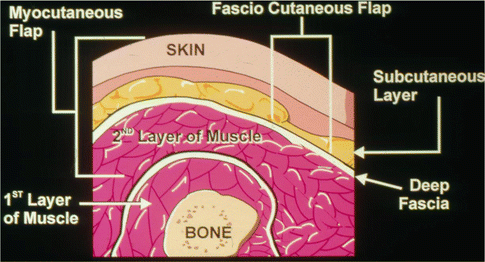
Cutaneous (skin only); fasciocutaneous, meaning skin and the cutaneous layer with the deep fascia; muscle only; or muscular-cutaneous (Fig. 7.2).


Fig. 7.2
Diagram showing the principle of flap composition
3.
The blood supply to the flap can be random, axial (arterial pedicle), or free flap microsurgical (the artery and vein).
7.5 Methods and Strategies in Flap Selection
What type of flap should one utilize for a particular ulcer? The answer depends on the following factors that should be considered in flap selection:
1.
The patient’s primary disease, whether it is spinal injury, spina bifida, advanced neurological disease, or post geriatric disease, and whether the patient is confined to a wheelchair is considered. The surgeon should select a flap that will fill the ulcer defect and heal the wound in a short time. In addition to excellent skin surface and good padding over the bone, consideration of recurrence risk in certain types of patients requires leaving a reserve of sufficient skin and muscles, especially if the patient is in a young age group.
2.
In patients who are ambulatory with sensation, the selection of the flap should not impact the motor function of the patient in walking, climbing stairs, or flexion of the hip or knee. For example, the gluteus maximus muscle in its upper and lower portion should not be used as a rotation flap. When the gluteus muscle tendinous part is detached at the point of insertion, or in the case of the hamstring muscle advancement flap rotated from its base when transaction of the lower part of muscles is performed. The vastus lateralis muscle is a powerful component of the quadriceps mechanisms and should not be used in ambulatory patients. The motor defect of this muscle affects the flexion of the hip and extension of the knee joint. The appropriate (or suitable) flap in this circumstance is the fasciocutaneous flap from the thigh and gracilis muscle to repair ischial and perineal defects. For hip joint defects, the rectus abdominis or rectus femoris muscle is appropriate.
3.
For each anatomical ulcer location, there is a primary flap to be used for a primary virgin ulcer, that is, a first-time ulcer should be considered with attention to the primary disease of the patient. For example, for a sacrococcygeal ulcer, the gluteus maximus is used as a musculocutaneous flap in a rotation or island advancement flap. For an ischial ulcer, the lower portion of the gluteus maximus is used in a rotation flap or the V-Y hamstring is used in a musculocutaneous advancement flap, with or without the gracilis muscle. For a trochanteric ulcer, the tensor fascia lata is used in a V-Y advancement flap or a rotation form. In the case of a hip defect, the size of the defect or the existence of another ulcer determines the selection of the muscle, whether it is the vastus lateralis muscle flap or the rectus femoris muscle flap.
4.
In recurrent ulcers, the selection of flap is more complicated and depends on the local tissue available to be used. Taking into consideration the primary disease of the patient, the choice of fasciocutaneous flap or distant muscle flap depends on which primary muscle flaps have been used previously. For example, the vastus lateralis muscle flap is used to repair extensive recurrent ischioperineal ulcer. The author’s experience in these circumstances is to reuse previous flaps if possible (e.g., re-rotation of the gluteus maximus flap or re-advancement of the hamstring musculocutaneous flap). Each time these flaps are reused, the quality of skin and vascularity is affected and, consequently, healing is at risk. The surgeon should explain to the patient the risk of recurrence of ulceration and skin breakdown, causing depletion of skin and muscle reserve in the patient’s body. In raising flaps in recurrent ulceration, it is important to consider the vascularity of tissue dissected and whether the skin and muscle beneath will survive. It is often possible during surgery to see that the color of the skin is dull and dusky. To confirm this observation, a fluorescent dye is injected intravenously during surgery, and the perfusion of the tissue is observed by ultraviolet light, which shows a yellow coloration of the skin if it is fully perfused. Otherwise, the color of the skin is dark and dull when there is no blood perfusion.
7.6 Type of Anesthesia and Patient Positioning for Pressure Ulcer Surgery
7.6.1 Type of Anesthesia to Be Administered
Most spinal cord injury (SCI) patients do not have sensation from the chest down, some from the waist downward, which means that they do not have feeling over the surgical site. It may seem easy to perform surgery under intravenous sedation to keep the patient comfortable in a prone or lateral position, but in clinical practice it is not always safe, especially in tetraplegic patients with shoulder pain and limited movement. The important issue is the airway and breathing when the tetraplegic patient cannot breathe and expand their chest in a prone position secondary to the paralysis of the muscle of respiration. In addition, a large percentage of tetraplegic patients are sensitive to intravenous sedation, which depresses their respiration and makes them unresponsive to stimuli. It is unsafe to place tetraplegic or advanced neurologic patients with tracheostomy tube in a prone position and to administer intravenous sedation, which can carry a high risk to the patient. For these reasons, tetraplegic patients should have general anesthesia for flap surgery.
In the case of the paraplegic patient group or other neurological patients, the author’s experience in performing surgery under intravenous sedation has lead to the conclusion that there is a high risk from this practice. When the patient experiences discomfort and pain in the prone position, the anesthetist administers more intravenous sedation, which leads to the patient becoming unresponsive. The patient does not breathe well, resulting in low blood oxygenation. This has lead the author to change his practice to administration of general anesthesia for all patients. Another advantage of using general anesthesia is the monitoring of the patient that can be done by the anesthetist. A fast blood transfusion can be given if needed without discomfort to the patient, and extensive, prolonged surgery can be performed under general anesthesia. In tetraplegia (above the level of T-7), a patient can develop autonomic dysreflexia secondary to the stress of the surgery, which causes high blood pressure and can create extensive bleeding at the surgical field or bleeding in the brain. Control of this condition is manageable when the patient is under general anesthesia by administering intravenous nitroglycerine. In our practice, all patients are intubated in a supine position first and then turned to the prone position on the operating table over a chest roll. At the end of the surgery, the patient is turned in the supine position on the postoperative gurney while still intubated and then extubated safely.
7.6.2 Patient Position on the Operating Table
The classic location of pressure ulcers is on the posterior torso of the body, for example, the sacrococcygeal or ischioperineal ulcer. To have surgical access to these ulcers, the patient is placed in the prone position. When a patient has a trochanteric ulcer in addition to one of the ulcers mentioned above, the prone position is ideal for closing all the ulcers in one stage (Fig. 7.3). When a patient has only a trochanteric ulcer or hip infection, the lateral position is good. The patient can be supported in the lateral position by a bean bag; suctioning the air from the bag will convert it into a hard support for the patient. The author always places a foam pad between the patient and the bean bag to prevent any pressure from the hard bean bag (Fig. 7.4


Stay updated, free articles. Join our Telegram channel

Full access? Get Clinical Tree


