13 Forehead and temporal recontouring using calcium hydroxylapatite pre-mixed with lidocaine
Summary and Key Features
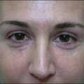
• Rejuvenation of the upper face should include correction of skin laxity at the temples and placement of volume in the forehead concavity
• Although multiple fillers are available to correct the upper third of the face, we recommend calcium hydroxylapatite for these sites specifically
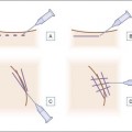
• Temple correction is best achieved through placement of boluses beginning at the temple and extending to the lateral canthus, followed by massage
• Forehead correction is achieved by placing a bolus of material into the inferior frontal eminence
• Postoperative edema is a common side effect that resolves spontaneously by 72 hours
• Relative ptosis of the eyebrows is also a temporary side effect and is a result of lidocaine and edema, typically resolving within 24 hours