Introduction of scar pathogenesis, epidemiology, and classifications
Indications and contraindications for laser scar treatment
Appropriate management of scars, including pre- and post-operative techniques
Potential adverse side effects of laser scar treatment
The use of laser therapy for atrophic scars, including ablative, nonablative and fractional resurfacing
Future directions of laser scar therapy
Introduction
The skin’s normal wound healing process involves coagulation, inflammation, proliferation and remodeling. Hypertrophic scars and keloids occur when the skin’s normal wound healing process goes away. Further, they more commonly occur in ethnic skin. There are multiple techniques used to treat these scars, and recent advances in laser therapy have made lasers a practical alternative. |
The skin is a primary barrier for protection and is often submitted to damage from surgical procedures, burns, trauma, infection, or inflammation. The skin has an incredible capacity to renew and repair itself through a process called wound healing, in which a cascade of mechanisms result in scarring.
The normal wound healing process, which results in a flat and flexible scar, can be divided into four major stages: coagulation, inflammation, proliferation, and remodeling. Once the injury to the blood vessels occurs, the inflammation phase begins after activating the clotting and complement cascade. Chemotactic factors such as prostaglandins, complement factors, and interleukins (i.e., IL-1) stimulate the migration of inflammatory cells such as neutrophils and monocytes/macrophages. These cells help wound debridement. Macrophages also release specific growth factors such as transforming growth factor Beta (TGF-β) and platelet-derived growth factors (PDGF), which initiate the formation of the provisional wound matrix. The proliferation phase involves the migration of fibroblasts, increased endothelial cells and keratinocytes to the site of injury. Fibroblasts are especially important in forming the extracellular matrix, which is made of collagens III and I, fibronectin, elastin, and proteoglycans. The keratinocytes start the re-epithelialization process with reconstruction of the disrupted basement membrane. Revascularization occurs with endothelial cells and angiogenic factors such as fibroblast growth factor (FGF). As maturation ensues, the collagen network and proteoglycans are also remodeled. While both collagen types III and I increase during the beginning of the wound healing process, the contribution of collagen type III decreases as remodeling takes place.
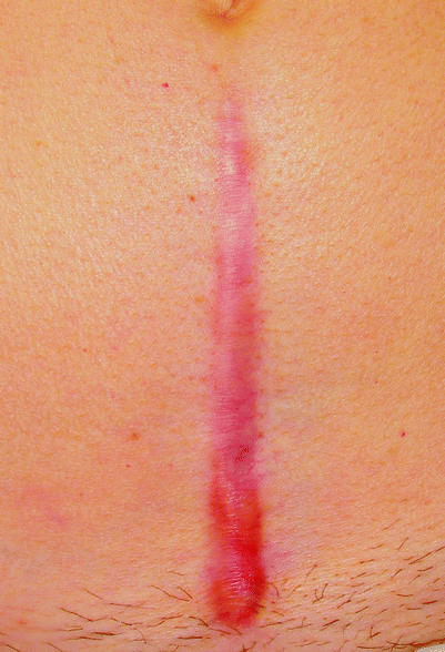
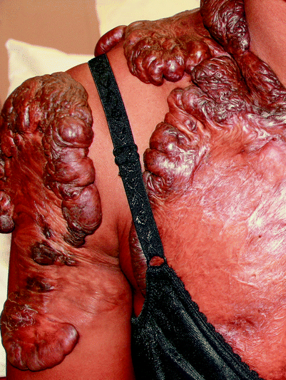
An abnormal response to the wound healing process results in hypertrophic scars (Fig. 1) and keloids (Fig. 2). These are essentially excess scar tissues that form at the site of injury. Fibroblasts from keloids abnormally respond by producing high levels of collagen, particularly type I. In contrast, fibroblasts from hypertrophic scars respond normally, with a slight increase in collagen. The collagen fibers in hypertrophic scars are more organized, while those in keloids are arranged in whorled, hyalinized bundles. Further, while angiogenesis diminishes during normal remodeling, but persists during development of keloids and hypertrophic scars.


Fig. 1
A well-circumscribed linear hypertrophic scar at lower abdomen after a gynecologic surgery

Fig. 2
Multiple keloids at the right arm, shoulder and chest wall of an African American woman
These can occur in all races, although they are more frequently found in ethnic skin, ranging from a 3–18 times higher incidence when compared with Caucasians. The incidence has been reported to be between 4.5% and 16% within African-Americans, Chinese, and Hispanics. Patients in their second to third decade of life are more commonly affected, with the same prevalence in both sexes.1–3
Hypertrophic scars are red, raised, and firm, and usually appear within 1 month at the injury site, especially sites under pressure or frequent movement. Keloids, which can often be disfiguring, are purple/red nodules that are often found beyond the original injury site. Unlike hypertrophic scars, these appear within weeks or even years from the initial injury. Keloids frequently are found on the earlobes, anterior chest, shoulders and upper back. Common processes that may lead to keloid formation are ear piercings, tattoos, infections, vaccination, burns, and inflammatory acne.
The treatment of hypertrophic scars and keloids is challenging due to high recurrence rates and adverse side effects. Previous methods for treatment have included surgery, cryotherapy, electrocautery and desiccation, dermabrasion, intralesional corticosteroids, 5-fluorouracil, and radiation. In terms of lasers, Ablative CO2 and Erbium:YAG lasers were used for hypertrophic scars but then discontinued. The pulsed dye laser (PDL) is currently the laser of choice for hypertrophic scars, which have yielded more successful results compared to keloids. It works via the principle of selective photothermolysis, in which the laser targets blood vessels, with the 585 nm wavelength specifically absorbed by hemoglobin. It is suggested that the microvascular destruction causes subsequent ischemia and reduction of scarring.
To appropriately treat hypertrophic scars and keloids, a thorough understanding of the processes by which they form is essential.
Indications and Contraindications
Indications for treatment include side effects, functional impairment, and aesthetics. It is important to take into account the patient’s skin type and lesion type prior to therapy. |
Patients typically seek medical help due to cosmetic issues or associated symptoms. Thus, indications for treatment of hypertrophic scars include functional impairment, cosmetic appearance, and pruritis and dysesthesias. The typical outcome of laser treatment is reduction of the redness and height of the scar, improvement of pliability, and symptomatic relief of pruritis.
Factors that need to be taken into consideration before treatment include patient skin type and lesion type. Fair skinned individuals (Fitzpatrick skin types I to III) tend to have a better response and fewer side effects to treatment. Patients with Fitzpatrick skin types IV to VI have an increased risk of laser light absorption by epidermal melanin, therefore less effective targeting of the skin and increased risk of postoperative hypo- or hyperpigmentation. It is suggested that in these patients, a test spot is done to predict any potential side effects. Lesion type also plays a role in the success of laser treatment. Lesions that have formed in less than a year and those that are red and raised have proved to be ideal for laser therapy.
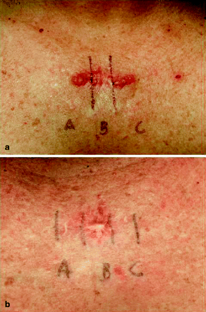
Lasers have been found to be useful as a preventative measure as well. The 585 nm PDL was found to be effective in preventing the formation of hypertrophic scars in burn wounds. Further, treating surgical scars starting on the day of suture removal has yielded optimal results (Figs. 3a and b).


Fig. 3
(a) A linear surgical scar on anterior mid upper chest. The right section received 585 nm PDL with pulse duration of 450 μs, the middle remained as a control, and the left section received 585 nm PDL with pulse duration of 1.5 ms. (b) One month after the third PDL treatment. There was a significant clinical difference between treated and untreated sites
Techniques
Pre-operative management includes obtaining a detailed history and exam of the patient’s scars, and if any prior therapy has been done. The area for treatment should be cleaned thoroughly and topical anesthetic may be used for comfort. It is important to take a picture of the scar before therapy to track its progress and note any side effects that may occur. Laser therapy involves appropriately calibrating and setting the laser parameters. It may be used alone or in conjunction with intralesional medical therapy. Multiple treatments are needed to obtain better results. Post-operatively, the patient should avoid the sun and keep the treatment area clean and protected. |
Pre-operative Management
It is important to get a good patient history before laser treatment. The history of the scar or keloid in terms of age, evolution, and previous treatments is helpful in determining treatment prognosis. It is ideal to treat hypertrophic scars early, possibly within the first few months of appearance. This is because of the numerous blood vessels present at this time. Previous treatments, such as cryotherapy, may cause increased fibrosis, and thus adjustments of laser parameters and treatment sessions may need to be made. Location of the scar is also important to note. Dierickx et al. have found that facial scars respond better to treatment.4 Nouri et al. agree and have found that facial, shoulder, and arm scars respond better than those on the anterior chest wall.5 However, Alster and Nanni found no relation between scar location and response to treatment.6
The PDL treatment for hypertrophic scars and keloids is an outpatient procedure. It typically does not require anesthesia unless requested by the patient. If so, a topical anesthetic cream may be applied 30–60 min before the procedure. Make-up any other creams must be removed from the treatment area with soap and water. In assessing the lesion, the physician should note its size, color, height, pliability, and associated symptoms. A picture should be taken prior to treatment for comparative purposes. It should be taken with the same camera, lighting, and distance. The patient and physician should be wearing eye protection at all times during the procedure.
Description of the Technique
The physician must first calibrate the laser and set parameters to be used. The ideal setting is 585 nm PDL at 450 μs pulse duration, using a spot size of 7–10 mm, with the fluence ranging from 3.5 to 7.5 J/cm2. When using a smaller spot size, the fluence should be increased; however, initial treatments should all begin with a lower fluence. In ethnic skin, we prefer using a lower fluence (3–3.5 J/cm2 with a 10 mm spot size) in order to reduce any complications from this procedure.
Once set, the physician should place the handpiece over one end of the scar and start applying the laser pulses over the entire scar surface in a continuous pattern until reaching the opposite end. A 10% overlap is generally accepted.
Laser treatment may be used alone or in combination therapy with intralesional corticosteroids or 5-FU. Alster compared PDL treatment alone with laser therapy combined with intralesional corticosteroid treatment and found that both were similarly effective with no significant difference.7 It is important to remember, however, that if using intralesional corticosteroids, the injections should be done following the laser procedure. If done before, blanching of the lesion occurs and there is substantial risk of losing the laser target. An average of 10–40 mg/mL immediately after laser treatment can be used.
Post-operative Management
Stay updated, free articles. Join our Telegram channel

Full access? Get Clinical Tree


