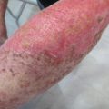
Fig. 1
Basal Cell carcinoma treated by open spray technique. In lager lesions,it is necessary to divide into smaller zones to promote more effective freezing and thawing cycles
The ideal treatment is considered repeated freeze-thaw cycles , rapid freezing, and slow thawing (the thaw time is usually two or three times longer than the freeze time) (Kuflik and Kuflik 2012; Lawrence and Tefler 2010).
Necrosis usually occurs at the center of the area of application, where the temperature should range between −30 °C and −40 °C. There is a rim of partially damaged tissue, and in the peripheral areas, some cells remain alive, but with such injury that triggers later apoptosis. Thermocouples inside the lesion or electrodes around can help to monitor the temperature (Pasquali et al. 2010; Kuflik and Kuflik 2012), but their placement is difficult to standardize (Petres et al. 1996). In practice, the temperature does not need to be measured because clinical studies suggest determined duration of freezing times for the most common skin lesions (Lawrence and Tefler 2010).
The lateral spread of freeze is also important and refers to the freezing of the tissue beyond the margins of the lesion. Benign lesions require usually 2–3 mm, and malignant lesions such as basal or squamous cell carcinomas should reach at least 3–5 mm, or more, if possible (Kuflik and Kuflik 2012).
Melanocytes are the most sensitive to freeze, with cell destruction at −4 °C to −7 °C (depigmentation may occur, especially in more pigmented patients). Keratinocyte death requires −20 to −30 °C. Fibroblasts are more resistant to freeze and require temperatures ranging between −30 °C and −35 °C to undergo cell death. Lower temperatures, such as −60 °C, are needed to destroy malignant lesions (Vujewich and Goldberg 2008; Pasqualli 2013). In general, benign conditions require more superficial freezing and, in fact, are better to undertreat a benign lesion than causes an anesthetic hypopigmentation or scar (Fig. 2).
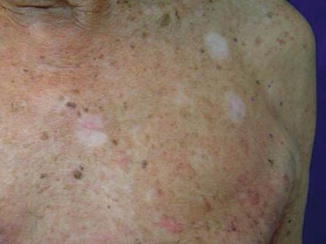
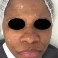
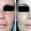
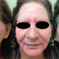
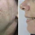
Fig. 2
Hypopigmentation after cryotherapy with liquid nitrogen for actinic keratosis treatment on the trunk
The conductivity of the material interposed between the lesion and the cryogen which determine the final freezing temperatures. Metals are ideal thermal conductors, such as copper (Pasquali et al. 2010).
Thick hyperkeratotic lesions have poor conductivity and should be debridated whenever possible, prior to cryogen application. It is also convenient to debulk nodular lesions or large tumoral masses, to avoid profuse bleeding.
Although there are several hypotheses, the full mechanism of tissue destruction by cryogens remains not completely understood (Petres et al. 1996).
Techniques and Equipment
Over the years, cryosurgical units have evolved from heavy bottles to highly efficient, low-weight, easy-to-use devices. There are storage tanks with 4, 5, 10, 25, 30, 35, and 50 liters of capacity for liquid nitrogen (Pasquali et al. 2010).
There are several techniques to perform cryosurgery. The choice of which should be used depending on the lesion and the operator preference:
Dipstick technique: The traditional dipping in a cotton-tipped applicator into a cup with liquid nitrogen is generally inadequate, because the freezing is slow and superficial. It is the oldest method (Kuflik and Kuflik 2012). It may be used in small verrucae or similar lesions (Petres et al. 1996).
Solidified carbon dioxide: A less commonly performed method. A crushed solidified carbon dioxide, contained in a disposable towel, is dipped in acetone and applied lightly onto the lesion, for mild freezing and exfoliation of the skin (“slush therapy”) (Kuflik and Kuflik 2012). It can be used for acne vulgaris, acne cysts, rosacea, and flat warts (Kuflik and Kuflik 2012).
Open spray: The most frequently used. Consists of a handheld cryosurgical unit with a fingertip trigger (Kuflik and Kuflik 2012; Vujewich and Goldberg 2008; Pasqualli 2013). There are different spray tips varying in size. Important factors in determining the amount of cold delivered to the lesion are the tip diameter, intermittent release of the nitrogen, and distance from tip to target (Pasquali et al. 2010). Longer spray times are required for thick and malignant lesions . Shorter times are reserved for benign, thin, and atrophic lesions (Vujewich and Goldberg 2008). Spraying can be delivered in an intermittent or in a continuous manner (Pasqualli 2013). The spray is directed to the lesion from a distance of 1–2 cm (Kuflik and Kuflik 2012). Superficial lesions require a 2–3-mm freeze margin, and malignant and deeper lesions should have a 5-mm margin (Vujewich and Goldberg 2008). The spray devices obtain a temperature of −40 °C to a depth of about 12 mm (Petres et al. 1996).
Confined spray/closed cone: A variation of the open technique, in which the liquid nitrogen is confined within a cone that is held against the skin. Plastic otoscope cones and specifically designed cones (polycarbonate) can be used.
Chamber: Another variation of the open technique. The spray is released through an orifice into a metal chamber, firmly held to the lesion. The turbulent movement of the nitrogen inside the chamber lowers the temperature even further. Lower temperatures are achieved faster and require extreme caution. It is usually limited to malignancies (Pasquali et al. 2010).
Close/cryoprobe/contact: A copper cryoprobe (precooled metal tip) is attached to the cryosurgical unit. The metal probe should be pressed against the lesion on the skin. It is useful for treating small and well-circumscribed lesions or in confined locations (Vujewich and Goldberg 2008).
The contact freezing achieves −40 °C of temperature but a depth of only about 4 mm (Petres et al. 1996).
Cryo Tweezers: A variation of the cryoprobe technique, successfully used in pedunculated lesions such as skin tags and warts (Usatine et al. 2015).
Intralesional : Ideal for voluminous or deep tumors. One or several sterile cannula probes are inserted into one side of the tumor and running it interstitially along the lesion (along the largest axis, at its deepest point) until it appears on the opposite side. Liquid nitrogen is sprayed into the cannula, and an ice cylinder is formed within the center of the lesion. There is minimal surface destruction compared to the previously mentioned techniques (Pasquali et al. 2010).
Preoperative Preparation/Patient Selection
A great advantage for cryosurgery is that it does not require a special surgical area (Pasquali et al. 2010). Therefore it is suitable for patients in wheelchairs or for those who cannot leave their home or nursery. It is a safe technique to patients with underlying medical conditions (heart diseases, bleeding disorders, metabolic diseases).
If there is any suspicion of malignancy, a skin biopsy needs to be performed prior to the cryotherapy procedure. Dermoscopy, ultrasound, radiography, and other imaging tests may be necessary. Once the location, type, and thickness of the lesion are well defined, the surgeon can decide which is the preferable technique to be used. (Pasquali et al. 2010).
Pearls:
Most lesions do not require previous treatment. In some cases, keratolytic substances, curettage, or debulking may be necessary.
Irregular surfaces are better treated with the open technique (spray).
Always work in bloodless areas. Blood increases the local temperature.
Vascular lesions are better treated with probes, such as hemangiomas and other vascular malformations (see “Laser Treatment of Vascular Lesions”).
Cartilage and bones are very resistant to freezing.
Local anesthesia, in most cases, is not required. It may be considered in very anxious patients and children (Pasquali et al. 2010) and in cases of deep freezing (chamber, probe, and intralesional techniques). Topical agents may help and should be applied 30–60 min before the procedure.
It is very important to inform, verbally and with a consent form, all the expected postoperative course, as possible side effects and probable cosmetic outcome (Pasquali et al. 2010; Pasqualli 2013; Usatine et al. 2015).
If using probes, be careful not to remove a probe stuck to the surface of the skin. (Pasquali et al. 2010) A small container with warm water may help in case a probe gets stuck to the skin (Pasqualli 2013).
Fractional cryosurgery may be helpful to avoid deformities and retractile scars in big lesions. It is performed in stages, first in the center of the lesion, reducing its size, and then repeated as necessary until the tumor diameter is smaller than 10 mm, at which the standard procedure is performed (Gonçalves 2009).
Contraindications
Inexperienced clinicians should avoid this procedure, which can do great harm if used inappropriately (Usatine et al. 2015).
Despite the lesions, some of them are better dealt with other treatments, such as suspected malignant invasive lesions (melanoma) (Usatine et al. 2015).
Morpheaform, infiltrative, micronodular, or recurrent basal cell carcinomas are rare, if ever indications for cryosurgery, such as poorly differentiated squamous cell carcinoma. If used, it would only be considered a palliative treatment (Usatine et al. 2015).
Inappropriate sites: Preauricular and nasolabial folds tend to have high recurrence rates; hair-bearing sites can evolute with permanent hair loss; Fitzpatrick’s skin type 4 or 5 patients should be averted about hypopigmentation or hyperpigmentation; lower leg tends to have a bad healing process, especially in patients with poor circulation (Usatine et al. 2015).
Cryotherapy Therapeutic Indications
There are several indications for cryotherapy in the dermatologic field. Above, we list all of them and detail the main and most common in the daily practice.
Benign Lesions (Electrosurgery)
Acne cysts
The advent of effective modern medication has decreased the use of cryosurgery to treat inflammatory acne. Some deep nodules may respond to freeze-thaw cycles of 10–20s. Superficial cysts require a single freeze of 5–10s, and the results can be dramatic. There may be temporary crusting. The results can be improved with intralesional triamcinolone (Usatine et al. 2015).
Acrochordons
Cryospray with a bent spray tip or a small aperture is a fast and easy method to treat this lesion. Cryo Tweezers is the least painful and efficient technique and especially useful in the eyelids (Usatine et al. 2015).
Angiofibromas
Angiofibromas or adenoma sebaceum are cutaneous lesions seen in tuberous sclerosis. Some case reports describe good results with repeated cycles of cryosurgery, but with long freeze times, that may evolute with hypopigmentation. Angiofibromas may be seen as smaller lesions in patients who do not have the syndrome and may respond well to cryospray or contact probe (Usatine et al. 2015).
Angiomas
Small vascular lesions such as senile angiomas (“cherry”) and spider angiomas may respond to cryosurgery using a cryoprobe, which compresses the lesion during freezing. Ten seconds should be needed. In cases of large angiomas, they may be anesthetized and shaved off, with electrosurgery of the base (Usatine et al. 2015).
Benign lichenoid keratosis
Also called lichen planus-like keratosis. Often needs a biopsy to be diagnosed. Once the diagnosis is defined, cryosurgery is an effective treatment option if there is any remaining lesion.
Chondrodermatitis nodularis helicis
It is a painful nodule on the pinna, related to mechanic pressure on the ear. It is crucial to be sure that the nodule is not a skin cancer before using a destructive method such as cryotherapy. When clearly benign, it can be frozen with a cryoprobe or spray for 10–20s. Additional treatment includes surgical excision and intralesional steroid injection (Usatine et al. 2015).
Viral warts/condyloma acuminate
Cryosurgery remains a standard treatment option to the treatment of viral warts in adults. Young children may not endure the pain (Usatine et al. 2015). The use of local anesthetic cream 1–2 h before therapy may be useful. Human papillomavirus lesions are usually sensitive to cryosurgical procedures and sometimes can be treated in one single session with excellent results (Pasquali et al. 2010).
As previously mentioned, reduce keratin of verrucae by shaving or using keratolytic substances may improve the results. Wet the area before the freeze cycle to increase cold conductivity.
In common warts, initially 1–2-mm halo ice should surround the wart and the ice field maintained for 5 s. Filiform warts can be treated with Cryo Tweezers. The maximum results can be achieved with 3 weeks of intervals sessions.
For the treatment of flat warts, cryosurgery must be carefully considered because there is a high risk of pigmentary changes.
Dermatofibroma
Using a freezing method, certainly the nodular component can be flattened, but a pale area may be left behind. Specific studies show that cryospray for at least 30s and a 2 mm border of freeze obtain a good or excellent result (Usatine et al. 2015). Some authors suggest 60s of cycles due to the fibrotic nature of the lesion (Vujewich and Goldberg 2008).Related posts:

Stay updated, free articles. Join our Telegram channel

Full access? Get Clinical Tree