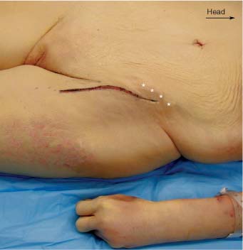
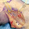
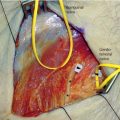
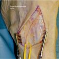
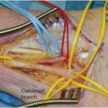
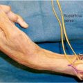
9 The femoral nerve takes its origin from the L2, L3, and L4 spinal nerve roots. It is the largest branch of the lumbar plexus and goes on to supply the iliacus muscles, the psoas muscle, the pectineus, the sartorius, and the muscles that make up the quadriceps femoris (rectus femoris, vastus lateralis, vastus intermedius, and vastus medialis). The iliopsoas muscle serves as the major flexor of the thigh, and the quadriceps muscles serve as the major extensor of the knee. The sartorius muscle serves to provide upward and rotatory motion of the thigh as the heel is raised to the opposite knee. The femoral nerve also supplies small articular branches to the hip and knee joint and to the adjacent vessels. Its sensory component supplies cutaneous branches to the anteromedial aspects of the thigh, leg, and medial foot. After the union of the anterior divisions of the second, third, and fourth lumbar spinal roots as the femoral nerve, the nerve passes inferolaterally in a retroperitoneal location, at first lying on the anterior surface of the psoas muscle. It then pierces through the psoas muscle to move to the medial edge of the muscle and then travels in the groove between the psoas and the iliacus. Entering into the thigh behind the inguinal ligament it lies lateral to the femoral vascular sheath in the femoral triangle. In moving from lateral to medial in the femoral triangle the structures encountered are nerve, artery, and vein, best remembered by the mnemonic NAV. Approximately 3.8 cm distal to the ligament it divides into multiple motor and sensory cutaneous branches. Motor branches are supplied to the muscles as previously named. Among the cutaneous sensory branches of the femoral nerve, the anterior femoral cutaneous nerve arises in the femoral triangle, pierces the fascia lata 8 to 10 cm distal to the inguinal ligament, and descends to knee level, supplying the skin and fascia over the front and medial sides of the thigh. Another branch, the saphenous nerve, is the largest and longest of the femoral branches. It arises at the femoral triangle and descends through it on the lateral side of the femoral vessels to enter the adductor canal. It crosses the vessels obliquely to lie on their medial side, anterior to the lower end of the adductor magnus. In the canal, branches of the saphenous communicate with branches of the anterior femoral cutaneous nerves to form the subsartorial plexus. At the lower end of the canal the saphenous nerve gives off an infrapatellar branch, which supplies sensation to the skin over the medial and anterior knee and the patellar ligament. The nerve continues down the medial aspect of the leg, pierces the fascia lata between the tendons of the sartorius and gracilis muscles, and gives off the sensory medial crural cutaneous branches to supply the skin of the medial leg. In the lower leg the saphenous nerve divides into its terminal branches, a smaller branch that follows the medial tibial border to the level of the ankle and a larger branch that supplies sensation to the medial side of the foot. When surgery on the very proximal or intrapelvic portion of the femoral nerve is required, the exposure can be challenging and may require the assistance of a general surgeon familiar with pelvic anatomy. There are several approaches to the proximal femoral nerve including an anterolateral extraperitoneal approach, a posterior extracavitary approach, and a transperitoneal approach.1,2 See Chapter 8 for exposure of the proximal femoral nerve in the pelvis. Exposure of the femoral nerve in the thigh is much more easily accomplished than the pelvic exposure. The patient is positioned supine with a small pillow or bolster under the ipsilateral knee to give a slight amount of flexion to the hip. This relaxes the sartorius muscle and facilitates retraction. As the nerve emerges, still within the pelvis, from between the psoas and iliacus muscles, at approximately the level of L5, it closely approximates the external iliac artery. Together with the artery it passes beneath the inguinal ligament and into the femoral triangle. The skin incision for exposure of the femoral nerve in the thigh is begun 2 cm below and parallel to the inguinal ligament. It extends laterally from the level of the anterior superior iliac spine, parallels the ligament, and then curves inferiorly at the medial third of the thigh. The incision should lie just lateral to the palpable pulse of the femoral artery (Fig. 9-1). The skin is infiltrated with a 1% lidocaine with epinephrine solution in a 1:100,000 concentration. When the skin and subcutaneous tissue are divided (Fig. 9-2), the fascia lata is uncovered and the outline of the sartorius muscle beneath it is visible (Fig. 9-3). The fascia lata should be divided parallel to the medial margin of the sartorius muscle and the sartorius muscle is then retracted laterally. The femoral nerve is invested with the iliacus fascia, which must be incised to expose the nerve (Fig. 9-4). Approximately 4 to 5 cm distal to the inguinal ligament the femoral nerve branches into several sensory and motor branches, and once the main nerve is identified these may be traced out as needed. Just medially the femoral artery is found (Fig. 9-5). The femoral vein is medial again to the artery, completing the anatomic mnemonic NAV that describes the position of the nerve, artery, and vein from lateral to medial (Fig. 9-6). For a more proximal exposure of the femoral nerve the inguinal ligament may be divided. Care must be taken to identify the superficial circumflex iliac artery as it crosses the path of the femoral nerve just before it emerges from beneath the inguinal ligament (Fig. 9-7). Once divided the femoral nerve is then visible lying atop the iliacus muscle, and the deep circumflex iliac artery is also seen crossing its path (Fig. 9-8). To expose the femoral nerve even more proximally, a muscle-splitting incision may be used whereby the external oblique and then the internal obliques are divided in turn above the inguinal ligament. This gives the exposure needed to retract the abdominal contents and expose the retroperitoneal space. Lateral to the femoral nerve the lateral femoral cutaneous nerve may be identified either proximally or distally as it traverses below the inguinal ligament (Fig. 9-9). Further dissection of the femoral nerve proximally, close to its formation at the level of the spine, requires an intrapelvic dissection as described in Chapter 8.
FEMORAL NERVE
ANATOMY
POSITIONING AND SURGICAL EXPOSURE
Femoral Nerve
Only gold members can continue reading. Log In or Register to continue

Full access? Get Clinical Tree


