Chapter 7. Facial Nerve Danger Zones
Nathaniel L. Holzman, MD; Sean T. Doherty, MD;
Brooke R. Seckel, MD, FACS
The most feared complication of facelift surgery is catastrophic injury to one of the major facial nerve branches. Injury can result in permanent facial deformity, numbness, dysethesia, and pain. Even a limited neurologic recovery can leave the patient with an undesirable aesthetic outcome, as well as discomfort. A thorough understanding of the nerve anatomy is paramount to avoiding injury. This chapter discusses the facial anatomic zones in which the major facial nerve branches are most susceptible to injury as described by Seckel (Fig. 7-1). Each of the seven sections highlights nerve origins, anatomic course, consequence of injury, and techniques for safe surgical dissection.
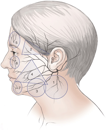
Figure 7-1 Side view highlighting the seven Facial Danger Zones.
ZONE 1
Facial danger zone 1 describes the region where the great auricular nerve is most susceptible to injury. With the patient’s head turned to the opposite side, a vertical line can be drawn, 6.5 cm in length, from the caudal edge of the external auditory canal. Centered around this point, a 3-cm radius circle is drawn. In this defined region, approximately 9 cm below the caudal edge of the external auditory canal, the great auricular nerve emerges at the posterior border of the sternocleidomastoid muscle, and becomes highly susceptible to injury, as it then runs superficial to the muscle belly. Injury to this nerve results in numbness or painful dysesthesia to the lower two-thirds of the ear, as well as the adjacent neck and cheek.
For safe dissection, after completing the postauricular incision, maintain a thin dissection plane just deep to the subcutaneuous fat, but superficial to the deep cervical fascia and sternocleidomastoid muscle. To identify the proper plane, retract the ear lobule forward and locate the two postauricular branches of the great auricular nerve that lie at the superior border of the dissection. Another tool for identifying the great auricular nerve is to visualize the external jugular vein throughout the dissection. Note the great auricular nerve will lie 0.5 to 1 cm posterior to this vessel. When plicating the platysmasuperficial musculoaponeurotic system (SMAS) layer, careful attention must be paid to avoid compression of the nerve. One way of avoiding a painful compressive neuropathy is to utilize the Hamra technique of tightening the platysma-SMAS layer via excision and repair of platysma bands anterior to the nerve.
ZONE 2
Danger zone 2 encompasses a triangular area through which the temporal branch of the facial nerve courses from its emergence beneath the parotid gland to the frontalis muscle. Laterally, a line is drawn from a point 0.5 cm inferior to the tragus to a point 2 cm superior to the lateral eyebrow. Inferiorly, a line is drawn along the zygoma to the lateral orbital rim, and medially, a third line connects the point 2 cm above the lateral eyebrow to the lateral orbital rim. It is here that the temporal branch of the facial nerve is most susceptible to injury when lying just below the temporoparietal fascia-SMAS layer. Nerve injury results in ipsilateral frontalis paralysis causing unilateral brow ptosis and asymmetric forehead animation.
Related posts:


Stay updated, free articles. Join our Telegram channel

Full access? Get Clinical Tree


