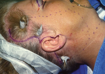
4 Stephen W. Perkins and Richard V. Balikian With the recent advances in surgical technique, a variety of anatomically sound techniques are available to the rhytidectomy surgeon. No one operation has been created that is best for treating the aging face. The astute surgeon will understand the importance of each patient’s individual needs and offer techniques most appropriate to each individual patient. One such technique that leads to a natural result is the superficial musculoaponeurotic system (SMAS) flap rhytidectomy. Proper incision planning and marking are crucial for creating long-term patient satisfaction. Avoiding changes in hairline and visible scars is critical to a successful outcome. Patients with well-hidden scars and unchanged hairlines will have more freedom with which to style their hair, leading to greater satisfaction. Often, the surgeon who takes care in incision planning will garner referrals by hairdressers and cosmeticians, who will recognize superior results.1 In considering the placement of incisions, three points must be considered: first, maintaining the preauricular tuft of hair, including the sideburn. Each patient is different in terms of the location of the lower portion of the sideburn and the width at which it extends anteriorly from the insertion of the helical curvature. If the preauricular tuft is 1 or 2 cm below the insertion of the superior portion of the helical insertion, it may be appropriate to design an incision that curves up into the temporal hair and allows some posterior superior lifting of the hairline. The curved hairline incision rather than a straight vertical incision is required to interrupt forces of contracture, maintain a minimally wide scar, and to avoid alopecia. As long as the hairline is not lifted higher than the insertion of the superior helical insertion, the patient will have no cosmetic disturbance of the area. If the sideburn is at the helical insertion preoperatively, an inferior sideburn incision is required. At no time should the incision be carried anteriorly around the sideburn tuft and along the pretemporal hairline. All scars in this area will be visible and cannot be camouflaged by the fine, severely sloped hair as it exits the skin naturally in a posterior direction. All incisions should be beveled to allow hair regrowth through scars created during incision.2 Second, preauricularly, the incision starts at the helical insertion and follows the apparent curvatures of the auricle itself to the helical root. The incision then goes behind the tragus ˜1 to 2 mm, then exits at the junction of the earlobe with the face. In male patients, a pretragal incision is used to avoid lifting and placing hair-bearing skin over the tragus (Fig. 4.1). Third, postauricularly, the incision must be directed up onto the posterior aspect of the auricle, above the sulcus so that when the ear settles posteriorly and the scar heals with some contracture of the skin, the scar falls into the sulcus. At the level of the helical insertion or eminence of the concha, the incisions curve gently toward the hairline. Depending on the amount of skin laxity needed to be removed from the neckline, the incision will be directed horizontally through the hairline (for minimal skin laxity) or down along the hairline (for greater skin laxity). When advancing the postauricular skin posteriorly and superiorly, the posterior hairline can then be approximated with no step-off or other deformity (Fig. 4.2).3 The beginning of the facelift operation requires treatment of the neck first prior to posterior shortening and suspension of the platysma muscle. In addition, treating the fatty tissues of the jowl, submentum, submandibular region, and neck sets the stage for proper contouring of the neck and jaw line with treatment of the SMAS tissues. The procedure is started by making a 2- to 3-cm incision in the submental crease, followed by 0.5-cm elevation of the skin to expose the subcutaneous tissue. After hemostasis is achieved, a small, 3-mm, round liposuction cannula with three rectangular holes on one side is used to preelevate tunnels into the jowl in a radial, fanlike fashion from the left sub-mandibular area completely to the submandibular area on the right. Once pretunneling has been accomplished, a judicious liposuction at 1 atm pressure of each jowl is performed to avoid dimpling. Symmetric and adequate liposuctioning can be accomplished by using the nondominant hand to palpate the jowls, then lifting the tissues and excess fat into the cannula. Care should be taken to avoid turning the holes of the cannula toward the dermis to avoid dimpling. For patients with significant lipoptosis, a 5-mm spatula liposuction cannula can be used in addition to the 3-mm cannula in the central neck compartment. Liposuction adds a tremendous amount to the overall initial and long-term results in facelifting with the caveat that one should underreduce the fat in any compartment rather than overremove it. Overzealous suctioning may make the ptotic submandibular gland more visible, thus a more difficult aesthetic issue for the patient.4 Fig. 4.1 Preauricular incision designed to prevent pretragal scarring and protection of the preauricular hair tuft. In most types of facelifting, further work is required to tighten the ptotic anterior platysma muscle as well as remove some subplatysmal fat in the anterior midline area. The Kelly clamp technique for submental platysmaplasty can be used for safe, reliable results. To accomplish the Kelly clamp platysmaplasty, complete undermining of the neck skin is required. This is done with Kahn beveled facelift dissection scissors. From the submental incision, the elevation is continued to the anterior part of the sternocleidomastoid bilaterally and across the cervicomental angle. Direct visualization of the remaining ptotic tissues is easily done. The forceps is used to pick up the loose anterior platysmal bands, as well as the subplatysmal fat that is redundant in the midline. A large curved Kelly clamp is then used to tighten these tissues in the anterior midline. Once tightened, creating firmness to the anterior tissues, sequential cauterization, excision, and suturing together with mattressing buried 3-0 Vicryl sutures (Ethicon, Somerville, NJ) is performed (Fig. 4.3
Extended Superficial Musculoaponeurotic System (SMAS) Facelift
♦ Preoperative Planning
♦ Surgical Technique
![]()
Stay updated, free articles. Join our Telegram channel


Full access? Get Clinical Tree


