Chapter 17. Extended SMAS Rhytidectomy
Bruce F. Connell, MD, FACS; Scott R. Miller, MD, FACS
INDICATIONS
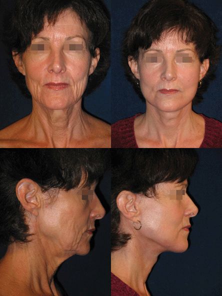
The indications for this method of rhytidectomy include patients with moderate to advanced facial aging, reflective of laxity of the soft tissues and their musculofascial support system, who desire top-quality, definitive facial rejuvenation with thoughtful tissue repositioning and discrete incisions (Fig. 17-1). The following is a basic technique, which is modified for each patient and for each side of the face, according to the specific anatomical problem.

Figure 17-1 Preoperative and 1-year postoperative frontal and lateral views following extended SMAS rhytidectomy. Proper use of the SMAS allows volume redistribution and restoration to a more youthful appearance. This procedure elevates the tissue as a unit, thereby providing sling support from the malar to the submental regions.
PREOPERATIVE PREPARATION
The patient’s medical status, prior operations and procedures, and results anticipated by both patient and surgeon should be reviewed. Each patient presents individual problems that require precise anatomical diagnosis, appropriate architectural planning, and skillfully executed repair. Without a thorough history, evaluation, and discussion of goals and expectations, surgery is ill-advised.
ANESTHESIA
Local infiltration, in combination with either full general anesthesia, total intravenous anesthesia (TIVA), or conscious sedation is appropriate for this procedure.
POSITION AND MARKINGS
The entire face and neck should be evaluated at rest and in animation. It is important to assess the skin redundancy with the shoulders down and with regard to quantity and vector of necessary removal. Abnormal fat accumulations, platysma laxity, and salivary gland and digastric muscle abnormalities are also marked.
DETAILS OF THE PROCEDURE
Incisions
The incisions are marked with attention to natural contour, texture, and skin tones. Respecting the hairline relative to the planned quantity and vector of skin shift determines incision at the sideburn margin versus in the temporal hair. The incision is then carried caudally at the anterior edge of the helical cartilage along the natural color change. It is passed deep into the depression superior to the tragus and then along the edge of the tragus. The V-shaped flap this creates upon inset and closure prevents scar contracture. The incision at the lower end of the tragus must be perpendicular to the long axis, thus defining the tragal height and preventing a subtragal skin fold. A planned incision 2 to 3 mm below the ear-cheek junction preserves the natural transition and avoids beard growth in an area difficult to shave.
With proper shift vectors, the postauricular scar does not descend. Consequently, the incision should be in the crease, not on the posterior surface of the ear. Extension posteriorly above or at the hairline depends on preoperative assessment of the amount and direction of neck skin to be removed. This is determined by pinching excessive skin with the patient seated during the preoperative examination. If incision at the hairline is chosen, a return behind the hairline at the fine hair at the nape of the neck with recipient site cut out and flap inset is necessary to maintain natural hair progression and prevent scar visibility. The submental incision, when necessary, is made posterior to the osseocutaneous adherence/crease. This allows release and redrape anteriorly, as well as mobility for exposure of fat, muscle, and/or glandular modifications.
Undermining
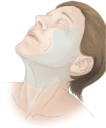
Skin flap elevation is to the malar eminence, more limited in the buccal region, and more extensive in the cervical region, and is based on skin laxity and crease release needs (Fig. 17-2).

Figure 17-2 Limited skin-flap elevation in buccal region maintains SMAS connections and vascular supply, allowing deep-layer support structurally and nutritionally.
Deep-Tissue Modifications
Stay updated, free articles. Join our Telegram channel

Full access? Get Clinical Tree


