Fig. 37.1
(a) Partly exposed tendon. (b) Half of the tendon is devascularised, the deep part remains in contact with the depth. Some vascularisation remains. (c) The whole tendon is completely detached. Vascularisation comes the edges, the risk of necrosis is high
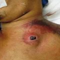
Stage 1: The paratenon is exposed and looks inert, dry and more or less hard. A few opening may be observed, giving access to the tendon itself. When properly managed, the paratenon may recover, be revascularised by the surrounding living structures.
Stage 2: The paratenon is lost and the tendon is directly exposed, some areas presenting a dead fleshy aspect, tendon fibres being easily dissociated with a single gauze. It is not easy to differentiate the still living areas from the dead parts, but the mechanical resistance to dissociation and hardness of the tendon fibres specifically differ (Fig. 37.2). Risks of complete loss of the tendon are important as infection of the exposed tendon may extend under the tendon sheets up and down and develop a large infected pocket. Modern management including negative pressure therapy after a surgical adapted debridement will enhance the progression of the granulation tissue coming from the edges and the deep part of the wound. The tendon will progressively be incorporated into this granulation tissue, and a coverage technique using either dermal substitute or flap will allow tendon salvage.
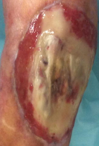
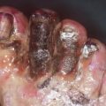
Fig. 37.2
Exposed infected tendon. Half of the structure is devascularised, soft and adherent. This portion should be debrided to the deep dense fibers
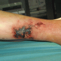
Stage 3: Clinically the tendon looks dry and hard having lost suppleness. No more connexion with the underlying structures is observed, the tendon lying inert over the wound (Figs. 37.3 and 37.4). Tunnels on each extremity are long and pus may be present. The tendon should be removed on the whole exposed area including the tunnels.
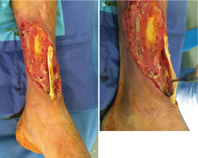
Fig. 37.3
(a) White pale devascularised tendon. (b) Adherence to the depth is lost. One can observe on the margins microvessels penetrating the deep part of the tendon
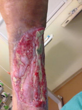
Fig. 37.4
Progressively the tendinous fibers are embedded into the granulation tissue. The surgical coverage may be realised using skin substitutes, skin grafts or flaps
When not managed properly complications of tendon exposure are multiple. The necrotic extension to the surrounding structure is often observed, infection may progress along the tendon sheet, issuing to secondary at distance infected wounds and complete destruction of the tendon and paratenon.
37.3 Modes of Treatment
37.3.1 Immobilisation
Immobilisation is a technique to be adapted to the limb segment and the structure to immobilise. Tendons being less rigid than bones, a strict tight immobilisation is not required. On the other hand, the course of a tendon of the foot being comprised between 2 and 4 cm, it is easy to understand that infection will tend to move with the tendon along the sheets and induce secondary infected zones involving the subcutaneous areas and the skin itself. In situations discovered lately, infection runs along the tendons or aponeurosis of the muscles and emerges as a secondary infection at the other extremity of a segment (seen in ischiatic pressure ulcers with a distal emergence of infection at the level of the knee).
37.3.2 Negative Pressure Wound Therapy
NPWT became a key factor to enhance tendon revascularisation and salvage. This technique represents a good alternative to flaps with less morbidity and more chances to save function, as the negative pressure acts as a regional booster for healing. Granulation tissue formation is increased around the tendon, providing a neovascularisation and preventing spreading of local infection. In an initial series of 16 patients, Lee et al. [1, 2] reported that NPWT facilitates the rapid formation of healthy granulation tissue on open wounds in the foot and ankle region and thus shortens healing time and minimises secondary soft tissue defect coverage procedures, reducing the need for a free flap to one single case. NPWT was also effective on infected patellar tendon salvage. Hong et al. [3] proposed an algorithm of decisions in soft tissue defects including NPWT.
37.3.3 Artificial Dermis
Several authors recently published clinical results concerning the use of artificial dermis in tendon coverage and more largely in soft tissue defects in the upper and lower extremities.
Attinger et al. [4] resume these possibilities as a step-by-step management of complex wounds with debridement, negative pressure wound therapy and coverage using dermal substitutes. These techniques are techniques described in chronic wounds, acute trauma wounds and burns [5, 6] with good results. Products used may either be double-layer dermis, mainly used in US, covered with a silicone film secondarily skin grafted, contrarily to one stage immediately covered with single layers proposed in Europe [7, 8] and Asia [9]. Other dermal substitutes have recently emerged on the market, aiming to the same objective which is to bring suppleness and prevent adherences of skin grafting to the underlying structures.
Dermal substitutes may also be used in the upper limbs [10] with good results in terms of mechanical possibilities of recovering skin capacities.
Related posts:


Stay updated, free articles. Join our Telegram channel

Full access? Get Clinical Tree


