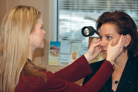
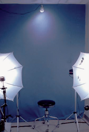
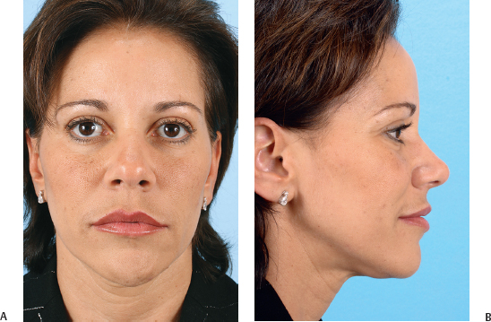
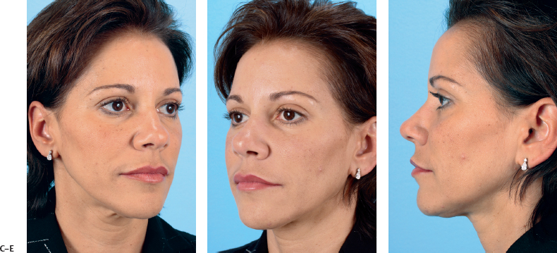
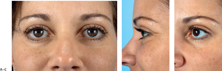
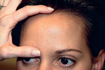
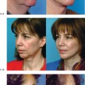
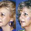
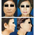
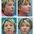
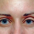
1 Ira D. Papel and Lisa E. Ishii Patients who present to a facial plastic surgeon for evaluation of an aging face represent a diverse population with a variety of different concerns. These patients may be male or female, range in age from their twenties to their seventies, and be from different ethnic backgrounds. They may have concerns about specific areas of their faces, or they may want the surgeon to recommend interventions that will improve their general attractiveness. They may have had previous cosmetic procedures or instead be presenting with a cosmetic concern for the very first time. Given the wide spectrum of patients presenting for aging face evaluation for the first time, it is advantageous for the facial plastic surgeon to follow a standard protocol with divergence as appropriate. The initial consultation should take place in the office or patient evaluation room (Fig. 1.1). Lighting should be appropriate for adequate physical examination. A mirror is typically necessary for patients to point out their specific concerns to the physician and for the physician to demonstrate proposed changes to the patient. Fig. 1.1 The initial evaluation takes place in an examination room with appropriate lighting. It is standard to photograph patients at the time of consultation after obtaining informed consent for photodocumentation. Patient photography should be standardized to enable comparison of preoperative and postoperative photos. Standardization is facilitated by photographing patients in a photo studio using a similar protocol for each photo session. Photographs should be taken in front of a consistent background with lighting that eliminates shadows without flattening the details of the face and skin. The common background color is light blue, which contrasts well with most natural skin tones. The modeling lights are typically umbrellas and or diffusers from a minimum of two directions (Fig. 1.2).1 Fig. 1.2 An appropriate photo studio. The typical photographic views for patients undergoing rhytidectomy are frontal, left and right profile, and left and right oblique (Fig. 1.3). Additional views may be added as appropriate given the procedure. For example, patients scheduled to undergo blepharoplasty should additionally have close-up views of the eyes in the open frontal, lateral, and oblique views (Fig. 1.4). The files can be uploaded for computer imaging with commercial systems available in a wide range of prices. Photographs viewed with such software can be altered to demonstrate proposed changes and can help to ensure that the patient and surgeon have similar expectations. In cases of facial rejuvenation surgery, computer imaging is useful for addressing the jawline, neck, and midface. Fig. 1.3 Typical photographic views for the rhytidectomy patient: (A) frontal and (B) right lateral. (C) Right oblique, (D) left oblique, and (E) left lateral. The evaluation should begin with a general physical assessment. Each patient should be considered in terms of overall body build and constitution, skin quality, hairline, and bone structure. Additionally, consider whether or not the patient has had prior surgery. Evaluate the face as a whole and in thirds, considering the upper, middle, and lower face and the neck separately. The initial evaluation should include an appraisal of facial proportions. The symmetric face when evaluated for width can be divided into equal vertical fifths. Each fifth should equal one intercanthal distance, the width of the neck should coincide with a line through the outer canthi on either side, and the lateral-most fifths extend from the lateral canthi to the lateral-most point of the helical rim.1 Vertical facial height should be measured from the trichion to the menton. Division into equal horizontal thirds should occur at the levels of the glabella and the subnasale. A separate facial height assessment considers only the middle third and lower third of the face. The middle third is delineated superiorly at the nasion with a division to the lower third at the subnasale with its further extension to the menton. In this method, the midfacial height should be 43% of the nasion to menton total distance with the lower facial height comprising the remaining 57%. The patient’s body mass index (BMI), calculated from height and weight measurements, should be considered prior to treatment planning. Patients with morbid obesity (BMI ≥35) have a three- to fourfold greater risk of respiratory depression from sedative drugs.2 Additionally, obesity is linked to systemic diseases known to elevate operative risk such as hypertension and diabetes.3 An increasing number of facial plastic surgical procedures are being performed in outpatient surgery centers as ambulatory surgery. The perioperative cardiopulmonary risk related to obesity or other medical disorders should be considered prior to scheduling outpatient surgery. Patients who are planning to lose weight should probably delay facial cosmetic surgery until after reaching their target weight. Loss of facial fat associated with overall body weight loss may result in additional skin laxity, particularly in the neck. This increased skin laxity will enable more accurate skin redraping during a facial rejuvenation procedure. Skin dyschromia, pore size, excess oil or dryness, and hairline position all contribute to the overall appearance of the aging face. Skin quality and hairline must be considered as part of the routine aging face evaluation for optimal treatment planning. Patients experiencing a facial aging appearance due to poor skin quality alone may benefit from a skin care regimen, even in the absence of surgery. For patients with poor skin quality who are planning to undergo a rejuvenating surgical procedure, a skin care regimen in combination with the surgical procedure will provide the greatest improvement in facial appearance. A long-term skin care program, including retinoids, toners, moisturization, and sun block protection, will enhance any surgical result. Fig. 1.5 Assessing the position of the anterior hairline. The position of the hairline must be considered when evaluating patients for surgical procedures and approaches (Fig. 1.5). Surgical procedures to rejuvenate the upper third of the face in patients with a high forehead or a receding hairline require careful planning to avoid conspicuous scars or abnormal hairlines postoperatively. For example, the incisions for an endoscopic brow lift may be placed in the hairline for a patient with a normal or low forehead but may need to be placed in a forehead crease for patients with a receding hairline.4 Frontal and peritemporal trichophytic incisions avoid anterior hairline elevation and maintain preoperative temporal tuft position. These incisions may be particularly useful in patients who have had previous procedures with hairline elevation or unfavorable repositioning of the temporal hair tuft.5
Evaluation of the Aging Face
♦ Physical Assessment
Body Build and Constitution
Skin Quality and Hairline
Bone Structure
Related posts:
Stay updated, free articles. Join our Telegram channel

Full access? Get Clinical Tree


