Chapter 11 Endoscopic Component Separation ![]()
1 Clinical Anatomy
 The rectus muscle can be fairly wide, up to 8 to 10 cm, and therefore the initial cut down for the balloon dissector must be performed in the lateral abdominal wall to avoid inadvertently placing the balloon in the rectus sheath.
 Understanding the anatomic characteristics of the external and internal oblique muscles is critical to ensuring accurate placement of the balloon dissector when performing an endoscopic component separation. The external oblique is primarily fascialike on the lower to midabdomen and is more muscular laterally and cephalad. The internal oblique is primarily muscular except for the most medial 2 to 3 cm of fascia just before its insertion into the linea semilunaris.
 Clearly identifying the linea semilunaris is important to safely performing a component separation. Inadvertently transecting the linea semilunaris during a component separation will result in a full thickness defect of the lateral abdominal wall and a troublesome hernia to repair.
2 Preoperative Considerations
2 Anatomic Considerations
 Skin Considerations
 Skin ulcerations and peristomal excoriations are carefully prepared preoperatively to maximize healing potential. Because skin flaps are not raised in this technique, skin preservation is very important.
 The primary advantage of an endoscopic component separation is the elimination of lipocutaneous skin flaps. This reduces wound infections and flap ischemia. In cases where excess skin must be resected or skin flaps will be necessary to mobilize the skin to the midline, the endoscopic component separation should not be used. In these cases an open component separation or concomitant panniculectomy is performed as described in other chapters.
 Musculofascial Considerations
 Ideal characteristics for an endoscopic component separation include a relatively wide, well-preserved rectus muscle. With a wide rectus muscle, a large mesh typically can be placed in the retrorectus position as an underlay (as described in Chapter 5 without a skin flap).
 Component separation techniques have limitations. Large defects, >20 cm in width, and multiple recurrent hernias with fixed noncompliant abdominal walls often cannot be medialized with this technique. In addition, patients with transverse incisions should not be approached with an endoscopic technique.
 Reconstructive Considerations
 The surgeon should consider whether skin flaps will be necessary to place the mesh. The absence of skin flaps can create technical challenges in placing a large sheet of mesh as an underlay. If appropriate mesh placement requires large skin flaps, I perform an open technique.
3 Operative Steps
1 Equipment
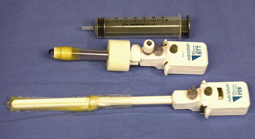
 Equipment needs include a10-mm, 30-degree laparoscope; bilateral inguinal hernia balloon dissector (Covidien, Norwalk, CT); 30-mL balloon-tipped trocar (Covidien, Norwalk, CT); laparoscopic trocars; and an ultrasonic dissector or LigaSure™ device (Covidien, Norwalk, CT) (Fig. 11-1).
 Patients receive appropriate preoperative antibiotics and invasive monitoring as needed, and epidural catheters are routinely placed for postoperative pain control.
 Trocar Strategy
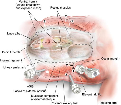
 Figure 11-2 shows trocar positioning, with lines showing the linea semilunaris, external oblique fascia, and costal margin
 The endoscopic component separation is typically performed first to avoid introducing any contamination from the midline wound into the lateral abdominal space. Given that these are closed spaces and no lymphatics are divided, drains are not routinely placed at the component separation sites.


