CHAPTER 22 Disorders of Pigmentation
Vitiligo
Vitiligo is a relatively common autoimmune disease of melanocytes, manifested by loss of pigment.
Epidemiology and clinical manifestations
Vitiligo is either most common or most recognized in darkly complexioned patients, particularly African Americans and those of Middle East extraction. This occurs in approximately 1% of patients1.
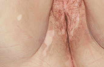
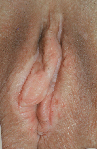
Vitiligo presents as well-demarcated, depigmented (totally white as compared to only light) patches (Figure 22.1). Vitiligo of the vulva usually affects keratinized, hair-bearing skin, the perineum, and perianal skin. Extragenital vitiligo is usually, but not always, present. A general skin examination most often shows similar patches over the extensor surfaces of joints, including the dorsal hands and fingers, wrists, knees, and elbows. Vitiligo is often perioroficial, occurring around the mouth, eyes, and nares. Vitiligo exhibits the Koebner phenomenon, in which areas of irritation or trauma are preferentially affected, partially accounting for this distribution (Figure 22.2).
Diagnosis and differential diagnosis
Vitiligo superficially resembles other white diseases, especially lichen sclerosus (Table 22.1). Lichen simplex chronicus in areas of moist modified mucous membranes is sometimes white as well. However, lichen simplex chronicus and lichen sclerosus show surface texture change, whereas vitiligo shows color change only.
| Diagnosis |
| Clinical appearance |
| Differential Diagnosis |
| Postinflammatory hypopigmentation |
| Lichen sclerosus |
| Management |
| Reassurance; therapy not usually successful |
| Topical corticosteroids twice daily may be useful |
| Tacrolimus or pimecrolimus twice daily can be tried |
Like vitiligo, postinflammatory hypopigmentation consists of light patches without texture change or thickening, but postinflammatory hypopigmentation is not white, only lighter in color than surrounding skin, and it is usually less well demarcated. Occasionally, inflammation, as a result of the Koebner phenomenon, precipitates vitiligo, so that depigmentation occurs in the area of active or past inflammatory disease (see Figure 22.2).
Pathogenesis
Vitiligo is disease that may be multifactorial, including an autoimmune factor, characterized by antibodies against melanocytes as well as cellular immunity1,2. Environmental factors and metabolic abnormalities leading to damage to melanocytes, particularly in the setting of oxidative stress, and possible neurologic abnormalities may also play roles3,4.
Therapy and prognosis
There is no predictably beneficial treatment for vitiligo. Other than reassurance and education, vitiligo of the genitalia is generally not treated. Occasionally, vitiligo improves with topical corticosteroid therapy, but this is most likely in children, and the experience of this author suggests that genital vitiligo is more difficult to treat successfully with topical corticosteroids than other areas5. There are reports of both benefit and no efficacy with the calcineurin inhibitors tacrolimus and pimecrolimus, although this is irritating to many patients6,7. Topical calcipotriene has been reported as useful in small series8. Although ultraviolet light treatments have been reported as beneficial for vitiligo, this therapy is usually avoided on genital skin because of the risk of squamous cell carcinoma9. The 308-nm excimer laser has been reported useful as well10. Finally, skin grafts sometimes prompt repigmentation of vitiligo.
Postinflammatory hypopigmentation
Postinflammatory hypopigmentation consists of lightening of the skin due to inflammation or injury. This is more common and more obvious in darkly pigmented races. Postinflammatory hypopigmentation consists of light patches of skin in the distribution of the inflammation or injury, and the degree of pigment loss is often proportional to the degree of injury (Figure 22.3). For example, cryotherapy can produce very white and permanent loss of pigment, whereas eczema most often results in poorly demarcated, transient mild lightening of the skin.
Lichen sclerosus (lichen sclerosus Et atrophicus, hypoplastic dystrophy): see Chapter 14
Lichen sclerosus is a well-recognized dermatosis characterized by white plaques and showing a predilection for the vulva. Probably multifactorial in origin with autoimmune mechanisms prominent, lichen sclerosus superficially resembles vitiligo. Classic lichen sclerosus presents as well-demarcated, white plaques encompassing the vulva, perineal body, and perianal skin (Figure 22.4). However, most patients describe itching and pain, and there is crinkling or thickening of the skin, unlike the asymptomatic white patches of vitiligo. Treatment consists of a potent topical corticosteroid and careful follow-up because of the slight increased risk for secondary squamous cell carcinoma.
Stay updated, free articles. Join our Telegram channel

Full access? Get Clinical Tree


