Chapter 18 Disorders of pigmentation
2. How do you diagnose a pigmentation disorder?
The clinical history is the most important aspect of the investigation of a pigmentation disorder. It should focus on the time of onset (such as at birth, during childhood, or later in life) and a family history. Other facts to be determined include any associated illness or symptoms, drug ingestion, chemical exposure, occupation, and exposure to sunlight, artificial ultraviolet light, heat, or ionizing radiation. Finally, a careful review of systems should be performed, followed by a skin examination.
3. What are the important elements of a skin examination of a patient with a pigmentation disorder?
The entire skin surface should be evaluated with attention to the color, shape, and distribution of the lesion(s). Lesion color helps to place the disorder into a specific category to aid in narrowing the diagnostic possibilities. The shape of a lesion is sometimes diagnostic. Linear areas of depigmentation, often in areas of trauma, are suggestive for vitiligo, whereas ash-leaf–shaped hypopigmented macules suggest tuberous sclerosis. Distribution of pigmentary changes also helps in diagnosis. Symmetrical depigmentation on the arms, legs, and/or torso suggests vitiligo. Increased pigmentation of the oral mucosa, axillae, and palmar creases is associated with Addison’s disease.
4. What is a Wood’s lamp?
A hand-held black light. A Wood’s lamp emits light in a narrow spectrum of long-wave ultraviolet to short-wave visible light. Hypopigmented areas appear lighter, and depigmented areas appear pure white. Furthermore, epidermal hyperpigmentation is enhanced (appears darker), whereas dermal hyperpigmentation is not enhanced.
Leukoderma: partial or complete loss of skin pigmentation
5. Name some heritable forms of leukoderma.
• Albinism is a group of autosomal recessive disorders characterized by generalized depigmented or hypopigmented skin and decreased visual acuity and nystagmus secondary to alterations in the formation of melanin.
• Waardenburg’s syndrome is an autosomal dominant disorder associated with congenital deafness, heterochromic irides, amelanotic skin macules, white forelock, laterally displaced medial canthi, and widening of the nasal root.
6. Name the skin disorder that manifests with complete loss of skin pigmentation.
Vitiligo is a depigmenting disorder due to loss of epidermal melanocytes. There are both familial and nonfamilial forms, and the overall incidence is 1% in the United States. Vitiligo has been reported to be associated with autoimmune disorders, including thyroid disease and diabetes mellitus type 1. The pathogenesis is not totally understood. Curiously enough, patients have both circulating antimelanocyte antibodies and skin-homing melanocyte-specific cytotoxic T lymphocytes. How these two portions of the immune system interact to result in melanocyte destruction is not understood. Vitiligo affects all races and affects both sexes equally.
Halder RM, Chappell JL: Vitiligo update, Semin Cutan Med Surg 28:86–92, 2009.
7. Describe the clinical appearance of the skin lesions in vitiligo.
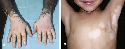
Typically, lesions of vitiligo are stark white with a well-demarcated border and no other skin changes. Sometimes, the border is hyperpigmented and rarely erythematous. Areas commonly affected are the periorbital, perioral, and anogenital areas, as well as the elbows, knees, axillae, inguinal folds, and forearms. Frequently, lesions of vitiligo develop symmetrically on the trunk and extremities (Fig. 18-1A). Vitiligo also causes depigmentation of hair (leukotrichia). Less commonly vitiligo is focal or segmental (Fig. 18-1B).
9. Do any factors influence the onset of vitiligo?
The patient presenting with vitiligo usually describes asymptomatic areas of skin that have rapidly lost all pigment. Rarely does the patient recall an associated illness, but skin trauma is commonly reported to cause vitiligo lesions. One caveat: Vitiligo occurs only in patients predisposed to the condition. Thus skin trauma will not induce vitiligo in nonpredisposed individuals.
10. Is vitiligo treatable?
Yes. Vitiligo repigments in small part from the border and mostly from the hair follicle. Localized vitiligo may be treated with high-potency topical steroids, topical tacrolimus and pimecrolimus, as well as topical calcipotriene. For more generalized vitiligo, narrow-band ultraviolet light B (UVB, 311 nm) is now the treatment of choice. It must be administered 2 to 3 times weekly for many months. All patients with vitiligo should use sunscreens to protect depigmented skin from sun damage.
Key Points: Racial Differences in Pigmentation
1. Pigmentation is determined by the type of melanin synthesized and the amount distributed to the surrounding keratinocytes.
2. Fair-skinned people produce a light brown form of melanin (pheomelanin), and distribute only small amounts to surrounding keratinocytes.
3. Melanocytes of darkly pigmented individuals produce a dark brown form of melanin (eumelanin), and distribute large amounts of it to neighboring keratinocytes.
Key Points: Pigmentation Disorders
2. Fortunately, the most common pigmentation disorders are benign, self-limited, and reversible. For example, one of the most common develops following a cutaneous inflammatory reaction, when the skin is either more darkly pigmented (postinflammatory hyperpigmentation) or less pigmented (postinflammatory hypopigmentation) than surrounding normal skin.
4. The two major types of cutaneous dyspigmentation are leukoderma and melanoderma. Patients with leukoderma present with areas of skin that appear lighter than surrounding normal skin, whereas patients with melanoderma have skin that appears darker than normal.
Key Points: Sun-tanning
1. Sunlight stimulates human epidermal melanocytes to increase melanin synthesis and stimulates increased melanocyte transfer of melanosomes to keratinocytes. This melanocyte response to sunlight is called tanning.
2. The action spectrum of sunlight that causes tanning is the ultraviolet spectrum (wavelengths 290 to 400 nm).
3. Excess sunlight exposure causes abnormal melanocyte function, resulting in areas of melanocyte overproduction of melanin and increased melanocyte proliferation.
4. Overproduction of melanin in a localized area causes the development of brown macules called freckles.
11. What is piebaldism?
Piebaldism is an uncommon autosomal dominant depigmentation disorder that is characterized by a white scalp forelock and hyperpigmented macules within areas of skin depigmentation. Piebaldism is due to mutations on the KIT protooncogene that is located on chromosome 4. A normal KIT receptor is required for normal development and migration of melanocytes. Melanocytes migrate during embryologic development in a dorsal-to-ventral direction; melanocytes in piebaldism fail to properly migrate to ventral skin surfaces, such as the forehead, abdomen, and volar arms and legs. For this reason, depigmented areas in piebaldism predominate on ventral skin surfaces. Patients are otherwise healthy. There is no treatment available for piebaldism.
12. What is albinism?
Albinism is a group of inherited disorders of the melanin pigment system. All forms are autosomal recessive. In oculocutaneous albinism type 1 (OCA1), there is a defect in the enzyme tyrosinase with an absence in melanin synthesis. Generally, albinism presents as depigmented or hypopigmented skin and hair, nystagmus, photophobia, and decreased visual acuity (Fig. 18-2


Stay updated, free articles. Join our Telegram channel

Full access? Get Clinical Tree


