Fig. 3.1
ACL anatomy
Studies have demonstrated that most ACL reconstructions are performed by surgeons who perform fewer than 10–20 reconstructions per year [10]. Multiple studies report failure rates of reconstruction to be between 10 and 20 % [14–16]. In the United States, approximately 3,000–10,000 revision ACL reconstructions are performed annually [10, 17]. Approximately 15 % of all ACL reconstructions at specialized institutions are revisions.
Various factors influence the success or failure of primary ACL reconstructions including proper surgical technique, postoperative rehabilitation, injuries to meniscus, cartilage, and secondary restraints, and patient expectations. It is crucial to identify the cause of failure prior to revision ACL reconstruction to allow for detailed planning of the revision.
Definition of Failure in ACL Reconstruction
Despite the enormous advances in prevention, treatment, and rehabilitation after ACL injuries in recent years, there is no consensus for the exact definition of failure of ACL reconstruction [6].
The term “failure” is very broad and not specific, which renders the task of defining it rather difficult [18]. ACL failure can be defined by the inability to regain pre-injury function, recurrent instability, chronic pain, loss of range of motion, or osteoarthritis. Some even define failure as side-to-side difference of 4 mm or greater on the KT-1000 examination.
Countless definitions can be found in the literature [19]. The definition by Johnson and Coen et al. that is frequently used defines the failure of reconstruction of the ACL as the following: the presence of recurrent instability when performing daily or sports activities or a stable, but painful, knee, with more than 10° motion loss after the surgical procedure [20].
Based on this definition, some authors recommend revision surgery for patients with instability and symptomatic objective laxity after the primary reconstruction of the ligament. The objective criteria include KT-1000 with an anterior-posterior laxity greater than 5.5 mm when compared with the contralateral knee or the presence of a positive pivot shift test [21].
Reasons for Failure in Primary Reconstruction of ACL
Various factors have been suggested as possible reasons for considering a primary ACL reconstruction “failed.” It is critical to determine these factors prior to considering revision surgery.
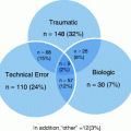
The reasons for failure can be generally divided into three groups: recurrent instability, postoperative complications (such as infections, stiffness, and arthritis), and persistent pain [6] (see Table 3.1).
Table 3.1
Reasons for failure in primary reconstruction of ACL |
|---|
Recurrent instability |
Postoperative complications (such as infections, rigidity, and arthritis) |
Persistent pain |
Recurrent Instability
Recurrent instability is defined as the inability to restore stability in the sagittal and rotational plane after reconstruction of the ACL, leading to patient complaints [6].
Instability after reconstruction may be due to trauma, technical error, failure of initial diagnosis, early return to sport, or inadequate postoperative rehabilitation and failure of graft incorporation [14, 22, 23].
Technical errors have been shown to contribute to the failure of ACL reconstruction in 22–79 % of cases. This group includes improper positioning of the tunnels, improper graft choice, and failure to diagnose conditions associated with the ACL rupture [24–27].
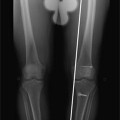
Poor positioning of the femoral tunnel is the most common technical error and has been documented as the cause for failure of ACL reconstruction in 36 % of cases (see Fig. 3.2). In a series of cases, Van Dijck et al. demonstrated that improper positioning of the graft results in a greater number of meniscal and cyclops lesions after the primary ACL reconstruction [28].
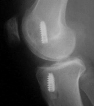
Fig. 3.2
Anterior position of the femoral tunnel
Failure to diagnose conditions associated with ACL rupture also plays a fundamental role. Missed medial or lateral side laxity and/or malalignment of the lower limb can also contribute to graft failure. The presence of these factors can produce an increase in loads and stresses placed on the graft during the postoperative period, leading to laxity and early failure [6, 29, 30].
Postoperative Complications
The loss of range of motion is considered to be the most common cause for unsatisfactory outcomes after ACL reconstruction in many studies [33, 34]. Various factors have been described as possible causes for this complication, such as poor positioning of the graft, a prolonged period of immobilization, arthrofibrosis, excessive tension of the graft, cyclops, persistent pain, and the time between injury and surgery. The loss of extension is more common than the loss of flexion [33, 34]. The cause of this complication should be determined and, if possible, corrected before the revision surgery. The objective of treatment is an improved range of motion and knee function, which can be achieved through rehabilitation exercises and/or open or arthroscopic debridement of the knee.
The presence of persistent pain after ACL reconstruction can arise from various factors, including chondral injuries, meniscal injuries, neuroma pain from the graft harvest site, arthritis, patellofemoral pain, bruises of the bone that occur during initial trauma, and synovitis. However, the diagnosis in some situations is complex due to the multifactorial nature of the problem and the difficulty in differentiating the symptoms of recurrent instability and persistent pain reported by the patient [35, 36].
Diagnosis
History and Physical Exam
A detailed history is extremely important for the evaluation of a patient who is symptomatic after ACL reconstruction. It is through this history that most of the information will be collected and used for determining the possible cause of failure in the original surgery.
The mechanism of the injury reported by the patient can assist in the investigation of associated lesions and in determining the degree of energy of the trauma. Determining the time between injury and surgery, period of immobilization after the reconstruction and the presurgery range of motion can be useful in investigating a loss of motion [37]. Analysis of the medical records from the original reconstruction will provide information regarding the graft used, type of fixation, presence of associated lesions to cartilage/meniscus, whether a notchplasty was performed, and the physical examination under anesthesia. Other important information includes the rehabilitation program and possible postoperative complications, as well as time to and level of return to sports. With respect to return to sport, one should investigate if the expectations were realistic and if new trauma occurred following the original reconstruction [38]. Previous imagings, such as X-rays, magnetic resonance images, and arthroscopy photos, are also useful.
The physical examination of the patient’s knee should be conducted after a complete history. During inspection, one should observe the alignment of the lower extremities in the sagittal and coronal planes. The presence of varus and/or valgus alignment of the lower extremity is important to evaluate the cause of failure of the original reconstruction and is fundamental to planning revision surgery. The presence of contractures in flexion or extension should also be noted. The presence of healed incisions about the knee provides clues on the type of graft and the fixation used in the previous surgery. The musculature of the lower limbs should be observed for atrophy. Gait should be evaluated for varus or valgus thrust due to ligamentous laxity [6]. Palpation of various anatomical structures of the knee should be performed to evaluate for painful areas and crepitation during flexion and extension of the knee.
The three physical exam maneuvers to evaluate the ACL are the Lachman test, the anterior drawer test, and the pivot shift test. A meta-analysis of studies focused on the clinical diagnosis of ACL rupture demonstrated that the Lachman test is the most sensitive and the pivot shift test the most specific, with both being indicated when there is a suspicion of rupture and/or re-rupture of the ACL [39].
The Lachman test performed in the supine position with the knee in semi-flexion is classified as grade 1 (0–5 mm), grade 2 (6–10 mm), and grade 3 (>10 mm) when compared with the healthy contralateral knee [6] (see Fig. 3.3).
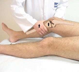
Fig. 3.3
Lachman test
The presence of previous injury to the ACL in the contralateral knee makes the evaluation more difficult. In cases where there is suspected rupture of the graft after ACL reconstruction and a previous injury to the contralateral knee, the use of records from previous evaluations is extremely valuable. Recently, Mulligan et al. investigated the utility and accuracy of the Lachman test when performed in the prone position to facilitate the test and proper stability of the thigh for larger patients and examiners with small hands. As the sensitivity and specificity of this test was 70 % and 97 %, respectively, it was concluded that the Lachman test performed in the prone position was a good alternative for examining patients with a suspected ACL injury when considered with other diagnostic criteria [40].
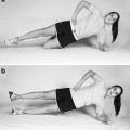
The anterior drawer test is performed with the patient in the supine position with the hip flexed to 45° and the knee flexed to 90° (see Fig. 3.4). It is graded according to the International Knee Documentation Committee [41] as normal (0–2 mm), nearly normal (3–5 mm), abnormal (6–10 mm), or severely abnormal (>10 mm), with the measurements based on the anterior translation of the tibia when compared with the contralateral knee [39]. This test should be performed with the patient’s leg at neutral, internal, and external rotation, thus allowing an evaluation of both antero-posterior instability and the medial and lateral compartments.
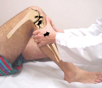
Fig. 3.4
Anterior drawer test
The pivot shift test is the most specific test for diagnosis of ACL injury [39]. This test is graded, according to the IKDC [41], as normal, glide (+), clunk (++), or gross (+++) [6]. The pivot shift test evaluates the presence of rotational instability that may occur, for example, in sports with quick changes in direction (see Fig. 3.5). Kocher et al. demonstrated that the Lachman test does not show a positive correlation with subjective evaluations after reconstruction of the ACL [42].
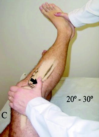
Fig. 3.5
Pivot shift test
In the same study, the pivot shift test had a positive correlation with patient satisfaction, global function of the knee, and return to sports after reconstruction of the ACL [42]. However, the sensitivity of this test is influenced by various factors, such as the speed with which the maneuver is performed, the angle of hip abduction during the test, and the magnitude of the force applied by the examiner during the test [43].
Thus, in recent years, various authors have investigated options for objectively quantifying rotational instability of the knee [44–46]. Most of these were performed with the assistance of computerized navigation. Some authors even published studies demonstrating a high accuracy and agreement of these evaluation methods [45–47].
At present, most orthopedic surgeons use measurements of the antero-posterior translation of the tibia (KT1000/2000) and questionnaires on satisfaction and function of the knee to evaluate the stability of the joint and the success of the ACL reconstruction [48]. However, these evaluation methods do not necessarily guarantee that knee function was totally restored [49]. Moreover, they do not necessarily provide information with regard to the level of control and dynamic rotational stability. Thus, in future years, we should improve and develop objective systems for evaluating the rotational instability of the knee to better evaluate our patients.
For an objective evaluation of the isolated antero-posterior translation of the tibia, advances are already being made in terms of improvement in the various systems for measurement. The most commonly used system is the KT-1000/2000, which is accepted worldwide as a tool for measuring the antero-posterior translation of the tibia (see Fig. 3.6). The KT 1000/2000 has a high level of accuracy, even when compared with more precise systems of evaluation, such as computerized navigation [50]. Previous studies showed that the normal difference between measurements of the anterior translation of the tibia between the knees generally does not exceed 3 mm; a difference of 3–5 mm is considered to indicate a partial loss of ACL function, and a difference >5 mm is considered to indicate a total loss of function of the ligament [51].
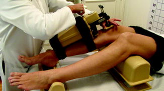
Fig. 3.6
KT-1000
Examination of the lateral and medial ligament complexes is essential prior to considering revision surgery. Failure to diagnose and treat associated ligament laxity can produce an increase in the load on the graft and subsequent graft failure. Injuries to the postero-lateral corner of the knee are the most commonly untreated injuries, and have been reported in 10–15 % of patients with chronic insufficiency of the ACL [29]. Other structures, such as the medial collateral ligament, the posterior horn of the medial meniscus and the capsule, play an important role in the stability of the knee in ACL injuries and should be investigated thoroughly in the clinical examination of these patients [52


Related posts:

Stay updated, free articles. Join our Telegram channel
Full access? Get Clinical Tree
