Chapter 14. Deep Plane Rhytidectomy
Samuel J. Lin, MD, FACS; Olubimpe A. Ayeni, MD, MPH, FRCSC; Thomas A. Mustoe, MD
INDICATIONS
The indications for this method of rhytidectomy include the patient with adequate expectations and anatomy for facial and neck rejuvenation. The advantages of this procedure are the ability to rejuvenate the midface and mouth area, as well as the neck.
PREOPERATIVE PREPARATION
Adequate time in consultation and preoperative planning is essential. The patient’s expectations must be explored. An older patient with significant laxity will have an incomplete correction at one year as a result of inevitable tissue relaxation. A history of prior rhytidectomy is important for technical planning prior to performing revision rhytidectomy.
ANESTHESIA
Either monitored conscious sedation or general anesthesia is suitable for this procedure.
POSITION/MARKINGS
The patient is placed in the supine position, prepped and draped in the usual sterile fashion for head and neck surgery. The patient’s head may be placed on a doughnut.
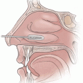
The blue outline signifies the extent of the superficial musculoaponeurotic system (SMAS) layer (Fig. 14-1A). Although variations may be present, standard rhytidectomy incisions (Fig. 14-1B) include a preauricular component with an anterior incision that borders the sideburn, with a V shape to allow correction of unequal incision length without a dog-ear. If more skin excision is anticipated, it useful to extend the incision up into the temporal scalp as well, which allows some posterior movement of the upper midface skin. The preauricular incision may be selected in a natural appearing crease; alternatively, the incision may extend behind the tragal cartilage. The latter is generally preferable except in secondary facelifts.
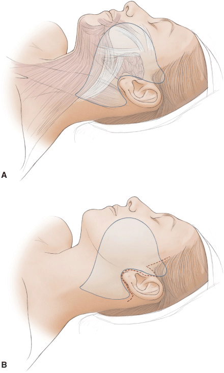
Figure 14-1 A. Outline of SMAS. B. Standard rhytidectomy incision.
INCISION AND EXPOSURE
A large volume of dilute local anesthesia is useful (100 mL 1% Xylocaine, 250 mL saline), which helps in defining the tissue planes. Following the injection of local vasoconstrictive/anesthetic agents, the incision is made with a no. 15 blade. Using sharp dissection, the incision is deepened through the subcutaneous tissues. Above the zygomatic arch, a superficial dissection is carried out. The SMAS is incised at the level of the arch, and 1 cm anterior to the ear. Above the arch, gentle blunt dissection with a peanut sponge may be used sparingly to separate the subcutaneous tissue from the temporoparietal fascia.
DETAILS OF THE PROCEDURE
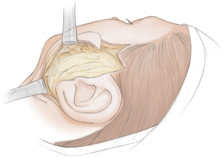
The preauricular dissection separates the overlying SMAS from the parotid fascia. Once the anterior border of the parotid is reached, blunt dissection can be utilized except when incising the retaining ligaments (Fig. 14-2). The postauricular incision is also deepened and dissection is performed sharply with a knife to raise the superficial layer of the deep cervical fascia off the sternocleidomastoid fascia from a superior to inferior direction. A zone of adherence exists at the posterior border of the SMAS, which is just anterior to the greater auricular nerve (it is routinely seen and identified in this dissection), and so dissection proceeds carefully in this critical area to join the posterior dissection and the anterior dissection.
Related posts:


Stay updated, free articles. Join our Telegram channel

Full access? Get Clinical Tree


