Chapter 35 Cutaneous manifestations of internal malignancy
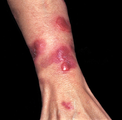
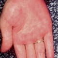
Figure 35-1. Sweet’s syndrome demonstrating painful red indurated plaques on on the hand and arm.
(Courtesy of James E. Fitzpatrick, MD.)
Bae-Harboe Y-SC, Salter SA, Kimball A: Acute febrile neutrophilic dermatosis, eMedicine Online August 2009. Available at: http://www.emedicine.com.
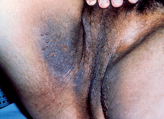
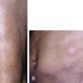
Figure 35-2. Acanthosis nigricans with hyperpigmented velvety skin lesions and small tags on the proximal thigh and groin.
Miller J, Rapini R: Acanthosis nigricans, eMedicine Online, June 2002. Available at: http://www.emedicine.com/derm/topic1.htm.
Zettouni N, Harvey N: Glucagonoma syndrome, eMedicine Online, http://www.emedicine.com. May 2008.
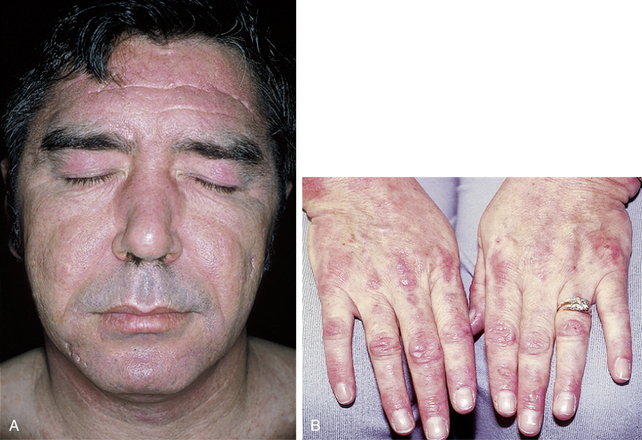
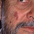
In addition to the facial rash, lesions on the scalp, neck, upper trunk, and extensor extremities are common. As the lesions mature, scaling and atrophy may develop. The erythema on the hands occurs over the knuckles rather than over the phalanges, as is typical of lupus erythematosus. Cuticular telangiectasias can be seen in both lupus erythematosus and dermatomyositis. Frequently, flat-topped, red-to-violaceous papules known as Gottron’s papules develop over the knuckles of patients with dermatomyositis (Fig. 35-4B).
Stay updated, free articles. Join our Telegram channel

Full access? Get Clinical Tree


