Fig. 45.1
(a, b) Preoperative view and preoperative drawings. The 58-year-old patient had a multicentric intraductal carcinoma in situ in the right breast. The breast was of large size and ptotic. Drawings were made for a skin-reducing, skin-sparing mastectomy
45.2 Surgery
A skin-reducing mastectomy with resection of the areola was performed with a resection weight of 810 g. Intraoperative sentinel node biopsy found two negative nodes. The pectoralis major muscle was dissected off its insertions in the inframammary fold and a 530 cc anatomical implant was inserted submuscularly. The distance between the dissected muscle and the inframammary fold was covered with a large extralight TiLOOP Bra mesh, which was fixed with resorbable sutures. Laterally the mesh was fixed to the fascia of the serratus anterior muscle. One drain was used and the wound closed in an inverted T fashion.
45.3 Clinical and Cosmetic Outcome
Initial postoperative course was uneventful. Final histology revealed a 120 mm intraductal carcinoma of high grade. The drainage was removed 10 days after surgery with less than 20 cc drainage fluid in 24 h.
Four weeks after surgery a small area of redness was noted in the lateral quadrant of the breast. Wound management was conservatively and antibiotics were given. The redness diminished in size, but following a couple of weeks, a 10 mm skin defect with the mesh penetrating through the skin was noticed. Bacterial culture from the fluid discharge was negative with no germs found. There were no signs of infection of the mesh or the underlying implant and nonsurgical wound management was done.
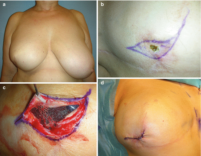
The defect gradually decreased in size, but no further progress in wound healing was seen over the next 7 months with the mesh penetrated through the mastectomy skin for a length of a couple of millimetres (Fig. 45.2a, b). The patient was scheduled for surgical revision with local excision of the fistula and the underlying mesh. The skin was closed after excessive mobilisation without tension (Fig. 45.2c, d). No drainage was used. Concomitantly an inferior pedicle-based reduction mammoplasty of the left breast was done for symmetrisation.

Get Clinical Tree app for offline access

Fig. 45.2
(a, b) Postoperative view after skin-reducing mastectomy and immediate implant reconstruction with a mesh (a). A small lesion with the mesh penetrating through the skin is visible in the lateral quadrant (b). (c, d


Stay updated, free articles. Join our Telegram channel

Full access? Get Clinical Tree


