Fig. 62.1
Preoperative view of breast asymmetry after two tumorectomies from the right breast and brachytherapy
As she had excessive tissue in the abdomen and was a healthy nonsmoker, we decided to excise the irradiated tissue in the right breast and to fill the defect with autologous tissue.
62.2 Surgery
Preoperatively the distance between the NAC and the clavicle was measured bilaterally, and a difference of 10 cm was confirmed (Fig. 62.2). A CT angiography was performed to determine the perforator vessels from the deep inferior epigastric artery (DIEA) to the skin in the lower abdomen (Blondeel 1999). They were marked on the skin using a Doppler probe. Intraoperatively a transverse incision was done above the nipple areola complex, and the scar tissue was removed. A DIEA perforator flap was harvested on one large perforator from the lower abdomen (Fig. 62.3). The flap was anastomosed microsurgically to the right internal mammary artery (Fig. 62.4). The width of the skin island was tailored to 11 cm in order to match the asymmetry of the position of the NACs.
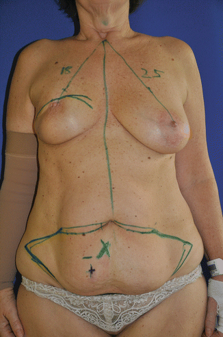
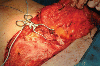
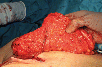

Fig. 62.2
Preoperative markings of the perforator vessels in the abdomen and the incision in the right breast

Fig. 62.3
Preparation of a large perforator vessel from the DIEA

Fig. 62.4
DIEA perforator flap anastomosed to the internal mammary vessels
62.3 Outcome
The postoperative course was uneventful. Early results showed some skin slough due to the badly perfused irradiated original breast skin (Fig. 62.5). Four months after the operation, good symmetry was achieved with additional improvement of the abdominal contour without sacrificing abdominal muscles (Fig. 62.6).
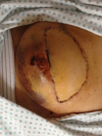
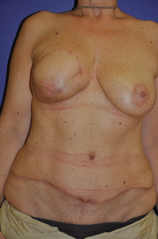

Fig. 62.5
Delayed healing of the irradiated skin envelope of the breast in the first postoperative period

Fig. 62.6
Symmetric breast size and position of the NACs 4 months postoperatively with improved contour of the abdominal wall
62.4 Comment of the Author
Stay updated, free articles. Join our Telegram channel

Full access? Get Clinical Tree


