Correction of Flatfoot Deformity in the Child

CHAPTER 17 Correction of Flatfoot Deformity in the Child


Pes planus is a common problem, and although the condition is predominantly idiopathic, in children it may be associated with neuromuscular disease and other disorders including tarsal coalition and the accessory navicular syndrome. The discussion in this chapter is limited to the flexible flatfoot, with or without the presence of an accessory navicular. By and large, similar principles of correction apply to treatment of flexible flatfoot in the child and to management of flatfoot in the adult, with the exception that a rupture of the posterior tibial tendon is not encountered in children, in whom rigid deformities are less common as well.


The difficulty in management of the flatfoot deformity in childhood is to know for whom and when treatment is required. Children who have severe flexible flatfeet, and who are symptomatic but have not responded well to management with orthoses, will require surgery. With those children who have severe flatfoot deformity but are asymptomatic, whether to persevere with nonoperative treatment or to proceed to surgical correction becomes a difficult decision. The adolescent with severe flatfoot who has a sibling or a parent with similar deformity that was not helped by orthoses also may benefit from earlier surgery. I find it helpful to examine the child every 6 months, to get a “feel” for the severity of the deformity and to look for any progression of the deformity or symptoms. There are of course children with asymptomatic flatfoot, whose parents are anxious about the shape of the feet, but who clearly do not require any treatment at all. With the passage of time, flexible feet in pediatric patients will become more rigid; this may happen in early adolescence or young adulthood. Adaptive changes inevitably take place in the hindfoot that alter its relationship with the forefoot. In order to keep the foot plantigrade, as the hindfoot everts and the calcaneus moves into valgus, the forefoot has to supinate. The Achilles tendon moves laterally with the calcaneus, and the axis of force on the subtalar joint changes, increasing the likelihood of a contracture of the gastrocnemius-soleus. As these structural changes take place, rigidity increases, and of course, the surgical treatment alternatives become bewilderingly complex.


In the young child with a symptomatic flexible flatfoot, I hope to reduce the hindfoot into neutral with a subtalar arthroereisis, with or without a lengthening of the gastrocnemius. If the forefoot is supinated with reduction of the hindfoot deformity, then it is useful to add a plantar flexion osteotomy of the medial cuneiform to maintain the forefoot in a plantigrade position. The many variations of this basic deformity must be appreciated; in some children, for example, the heel is in far more valgus, for which a subtalar arthroereisis does not provide sufficient correction, so a medial translational osteotomy of the calcaneus is required either as an isolated procedure or in addition to the arthroereisis. As the aforementioned adaptive changes take place, gradually increasing abduction of the forefoot relative to the hindfoot occurs, and the navicular moves off the head of the talus (uncovering of the talus). In these feet, a medial translational osteotomy of the calcaneus is not sufficient for correction, and a lengthening of the lateral column of the foot is required. Perhaps the most obvious difference in management of flatfoot between children and adults is that in the former, arthroereisis and osteotomy are emphasized, and arthrodesis should be avoided. Unfortunately, arthrodesis still has to be part of the treatment algorithm, because some adolescents will have a very rigid flatfoot, not associated with a tarsal coalition. Each of these procedures is discussed next.


ARTHROEREISIS


Indications and Rationale


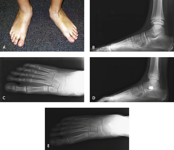
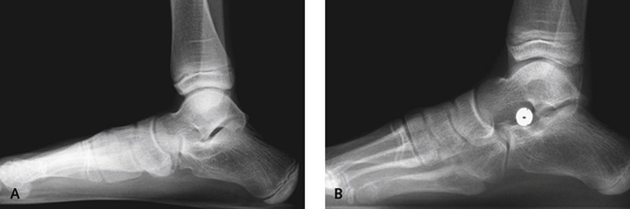
The goal of arthroereisis in the child is to properly orient the talus over the calcaneus; the joint is then allowed to remodel. This remodeling is expected to help prevent further problems later in life, such as degeneration or rigidity of the hindfoot. An arthroereisis implant can be considered to function as an internal orthotic device. This procedure has many advantages; most important, however, are the maintenance of motion it affords and the minimal associated morbidity. The indications for arthroereisis in the child are broad. Treatment results for children undergoing arthroereisis have been excellent, provided that the talonavicular joint is not significantly uncovered. The procedure seems to work very well in younger children who have predominantly heel valgus, presumably because they have more capacity for remodeling and adaptation of the forefoot (Figure 17-1). Once the talonavicular joint sags, particularly as seen on the lateral radiographic view, these feet seem to require more correction of the pronation deformity than a medial displacement calcaneal osteotomy can provide (Figure 17-2). If there is abduction deformity of the foot, with uncovering of the talonavicular joint, then neither the arthroereisis nor the medial displacement osteotomy is likely to be successful. In anecdotal reports, in the adult flatfoot, more of the talocalcaneal deformity is corrected with arthroereisis than with the abduction of the transverse tarsal joint. Most important, children are able to bear weight in a boot within days after the arthroereisis surgery.


The pediatric patient typically adapts to the arthroereisis very well, and the incidence of implant failure is low in this age-group. By contrast, in my own experience with use of arthroereisis as an adjunctive procedure in a group of carefully selected adult patients, sinus tarsi pain warranted implant retrieval in approximately half of the cases. In children, however, implant removal has been necessary in less than 10% of the cases, probably because the foot adapts as it matures.


One cause for failure of the implant regardless of the age of the patient is inadequate correction of the forefoot. When the hindfoot is restored to a neutral position with the implant, some supination of the forefoot occurs. If the forefoot is able to compensate by increased plantar flexion of the first metatarsal, then a plantigrade foot is maintained. If the supination exceeds this adaptive ability, however, then in order to maintain the forefoot in a plantigrade position, the hindfoot has to evert during the foot flat phase of gait. This increased eversion then compresses the subtalar implant, causing pain. For this reason, an opening wedge osteotomy of the medial cuneiform is necessary if supination is excessive.


Surgical Technique


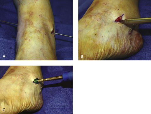
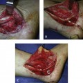
An incision is made in the sinus tarsi, approximately 1.5 cm in length. To locate the exact position for the incision, it is necessary to palpate the “soft spot” between the distal tip of the fibula and the anterior process of the calcaneus. The incision is placed inferior to the intermediate dorsal cutaneous branch of the superficial peroneal nerve and dorsal to the peroneal tendons. A guide pin that functions as a cannula for the arthroereisis dilators and sizers is inserted into the tarsal canal from lateral to medial, pushed through a puncture on the medial foot, and then clamped (Figure 17-3). The anatomy of the tarsal canal must be appreciated: The canal is shaped like an oblique cone and passes from anterolateral to posteromedial. The guide pin should therefore be inserted in the same plane as that of the tarsal canal and not directly medially. A slight resistance to the insertion of the pin can be felt as it traverses the interosseous ligament; then it is pushed through until it is protruding on the medial skin. The clamp on the guide pin prevents loss of position of the guide during repeated insertion of the sizers and trial implants.


Stay updated, free articles. Join our Telegram channel

Mar 6, 2016 | Posted by in Reconstructive surgery | Comments Off on Correction of Flatfoot Deformity in the Child

Full access? Get Clinical Tree

Get Clinical Tree app for offline access