Connective tissue diseases
The inflammatory disorders of connective tissue often affect several organs, as in systemic lupus erythematosus (LE), but they may also involve the skin alone (e.g. discoid LE). Autoantibodies are a feature of these diseases, which can thus be regarded as ‘autoimmune’.
Lupus erythematosus
Clinical presentation
Systemic lupus erythematosus (99% ANA, 50% Ro, 60% dsDNA positive)
Skin signs are found in 80%. The facial butterfly eruption (Fig. 1) is characteristic, but photosensitivity, discoid lesions, diffuse alopecia, mouth lesions and vasculitis also occur. Multisystem involvement with serological or haematological abnormalities must be demonstrated to diagnose systemic LE (Table 1). The female : male ratio is 8 : 1.
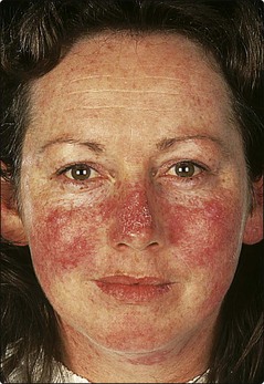
Table 1 Organ involvement in systemic LE
| Organ | Involvement |
|---|---|
| Skin | Photosensitivity, facial rash, vasculitis, hair loss, Raynaud’s phenomenon |
| Blood | Anaemia, thrombocytopenia |
| Joints | Arthritis, tenosynovitis, calcification |
| Kidney | Glomerulonephritis, nephrotic syndrome |
| Heart | Pericarditis, endocarditis, hypertension |
| Central nervous system | Psychosis, infarction, neuropathy |
| Lungs | Pneumonitis, effusion |
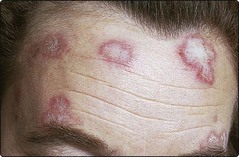
Discoid lupus erythematosus (35% ANA, 2% Ro, <5% dsDNA positive)
One or more round or oval plaques appear on the face, scalp or hands (Fig. 2). The lesions are well demarcated, red, atrophic, scaly and show keratin plugs in dilated follicles. Scarring leaves alopecia on the scalp and hypopigmentation in those with a pigmented skin. Remission occurs in over 50%. Internal involvement is not a feature, and only 6% develop systemic LE. Women outnumber men by 2 : 1.