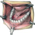
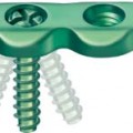
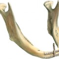
15 Condylar Neck Fractures: Delta-Shaped Plate and Endoscopic Approach Condylar fractures are frequent (Ellis et al., 1985, Silvennoinen et al., 1992), and there is growing evidence that their surgical treatment grants better results than conservative treatment (Worsaae and Thorn 1994, Eckelt et al., 2006). To minimize the risks of extraoral approaches—facial nerve damage, visible scars (Weinberg et al., 1995)—the endoscopically assisted intraoral approach has gained increasing attention. Endoscopically assisted condyle fracture reduction and osteosynthesis was introduced into the German literature by Fritzemeier and Bechthold (1993), then Mokros and Erle (1996). In the more international, English literature Lee’s group from San Francisco (Jacobovicz et al., 1998;Lee et al., 1998) were the first to describe this approach. Special devices to attach the endoscope and apply the osteosynthesis plates were developed by Lauer and Schmelzeisen (1999). Meanwhile the effectiveness of the technique has been demonstrated in several reports on a few cases as well as clinical follow-up studies (Chen et al., 1999; Kellman, 2004; Schön et al., 2005). Despite the progress in instrumentation technique and the increased popularity of this approach, open reduction with internal rigid fixation is still challenging, the reasons being: • confined space at the condylar neck • size and design of the osteosynthesis plate • biomechanical stability requiring two four-hole plates and eight screws To address and overcome these obstacles, the delta-shaped plate was designed with the gliding hole feature and four holes only. The biomechanical properties of the platehavebeen compared withother osteosynthesis systems in cadaver studies, and the clinical application of the plate has been tested in a recent follow-up study (Lauer et al., 2007a, b). The endoscopically assisted surgery requires a special set of instruments. In addition, it is helpful if the surgeon has some experience in treating subcondylar fractures openly and is used to performing orthognathic surgery. Besides the delta-shaped plate and the endoscope, additional instruments have proved to be helpful for performing this type of fracture surgery (Fig. 15.1), namely, two different types of low-profile right-angled drill and screwdriver, a drill guide, and a modified Schuchardt retractor with a suction and an endoscope attached. Further, it is particularly important to gain familiarity with the technique of performing surgery while visualizing the surgical site on a TV monitor. The monitor should be positioned so that direct viewing of the surgical field and the endoscopic picture are possible without turning the head. Particularly when starting the technique, simple fractures should be chosen. Low fractures with little displacement are ideal; for the more experienced surgeon all subcondylar and condylar neck fractures or fractures of the Spiessl and Schroll classification (Spiessl and Schroll, 1972) may be chosen. These authors differentiate six different types of fracture, of which Spiessl I–IV are approachable for repair with plates. Spiessl V and VI can be treated with screws and pins (see Chapters 17 and 18). Fresh fractures are best for surgery, when the initial swelling has settled. Delayed fracture repair makes what is already a technically demanding procedure, even more challenging. At the start of surgery, Ivy or Ernst ligatures are applied for intermaxillary fixation in the premolar region. The condyle fracture is exposed via one major and two additional small incisions (Fig. 15.2), which are referred to as “ports” in minimal invasive surgery. Most of the surgery is performed via the major port, and the additional ports facilitate the instrumentation. After infiltration with local anesthetics containing a vasoconstrictor (1 % lidocaine and epinephrine 1: 400 000), an S-shaped intraoral incision 3.5 cm long is made along the anterior border of the ramus of the mandible. To create the optical cavity for the endoscope between the mandibular ramus and the masseter muscle, careful dissection and elevation of the periosteum proceeds, avoiding disruption of the muscle (Fig. 15.3). Thus, troublesome bleeding and collapse of shredded muscle fibers into the cavity are avoided.
Introduction
Surgical Approach
Related posts:

Stay updated, free articles. Join our Telegram channel

Full access? Get Clinical Tree


