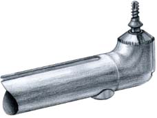
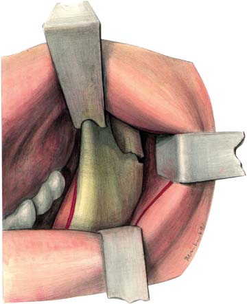
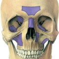
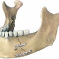
14 Condylar Neck Fracture Miniplates: Intraoral Approach Surgical treatment of condylar neck fractures is indicated where the dislocated proximal fragment is in such an unusual position that purely conservative treatment does not guarantee any success. Fig. 14.1 Right-angled drilling and screwdriving instrument. Fig. 14.2 Intraoral surgical approach to the condylar neck. The risk of damaging the facial nerve, which exists in open reduction via an extraoral approach, is avoidable when using intraoral access. This approach was first described by Silverman (1925) and later recommended by Steinhäuser (1964). Pape, Hauenstein, and Gerlach (1980) were the first to present a larger series with miniplate osteosynthesis of condylar neck fractures after an intra-oral approach. In a later follow-up, published by Horch, Gerlach, and Pape (1983), the poor results in some cases were explained by access difficulties; the approach was indicated only for low condylar fractures where the fracture line was running through the sigmoid notch. Later on, further recommendations for an intraoral approach were made by Jeter, van Sickels, and Nishioka (1988), Lachner, Clanton, and Waite (1991), Ellis and Dean (1993), Nehse and Maerker (1996), and Hochbahn et al. (1996). The difficulties previously described, involving access and control of the reduced condyle fragments, had been solved by using a right-angled drilling and screwdriving instrument (Fig. 14.1), and by the addition of endoscopic monitoring (Fritzemeier and Bechthold, 1993;Mokros and Erle, 1996; Gerlach, Mokros, and Erle, 1996). The condylar neck is approached through an incision over the anterior border of the ascending ramus extending into the lower buccal sulcus. The temporalis muscle is stripped from the anterior border and the masseter is reflected laterally by subperiostal dissection. Soft tissue reflection is enhanced by a special retractor placed at the dorsal border of the ramus. An additional notched retractor at the anterior border of the coronoid process allows good inspection of the sigmoid notch and the coronoid process (Figs. 14.2, 14.3
Introduction
Surgical Approach
![]()
Stay updated, free articles. Join our Telegram channel


Full access? Get Clinical Tree


