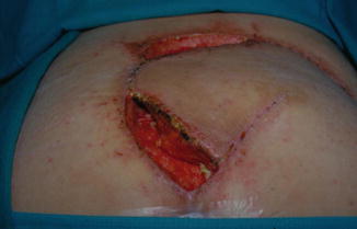
Fig. 15.1
Photograph of a patient in the prone position about 3 weeks after a gluteus maximus rotation flap was used to close a sacrococcygeal ulcer. A breakdown in the medial side of the flap can be seen, with some undermining as demonstrated by the Q-tip for about 3 cm under the flap. This indicates the formation of bursa under the flap

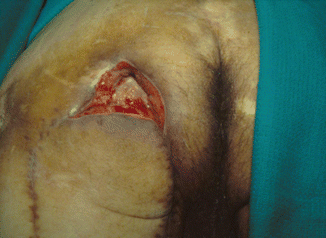
Fig. 15.2
Photograph of a patient in the prone position about 3 weeks after a gluteus maximus rotation flap was used to close a sacrococcygeal ulcer. Major skin necrosis and dehesion can be seen. The patient required a revision of his flap to close this open wound

Fig. 15.3
Photograph of a patient in the prone position about 3 weeks after a hamstring advancement flap. There is dehesion of the proximal part of the flap secondary to severe spasticity. This patient required revision of the flap with addition of a second flap, such as the gracilis muscle
15.4.1 Major Complications
Seroma is a frequent complication, which is detected after removal of the sutures when there is discharge leaking from the flap wound. Seroma development under the flap is secondary to bursal formation under the flap, which may result from severe uncontrolled spasticity, early removal of the drainage tube, a clog in the drainage tube caused by a clot, or when dead space under the flap is not closed completely. Management of this complication is aggressive irrigation of the space under the flap with normal saline, using a catheter inserted under the flap. The open area is then packed with a small strip of gauze soaked in normal saline or Dakin’s solution and changed twice per day. This management decreases the bacterial colonization of the space under the flap, which eventually will help to heal the open flap, and the flap will adhere back to its base. If conservative management is unsuccessful in closing the bursa, surgical management is indicated and entails opening part of the flap, debridement of all the granulating tissue or excising the bursa, and flap closure under a drainage system.
15.4.2 Wound Infection
The author’s experience is that wound infection is not a common complication, but it can occur when there are certain factors predisposing to infection. In 1992 [2], the author reported his experience in flap infection. In a 1-year period of time with a total of 76 flaps, 6 % became infected despite pre- and postoperative antibiotic coverage, adequate surgical excision and debridement of the ulcer, and aggressive intraoperative wound irrigation. The conclusion of the study showed that flaps close to the anus and perineum are more prone to develop infection than flaps in other areas of the body. Management is wound drainage and irrigation and wound packing with 0.25 % Dakin’s solution. Intravenous antibiotic is used if clinical signs of sepsis are present, such as fever or high white blood count.


