© Springer Japan 2016
Kenji Kabashima (ed.)Immunology of the Skin10.1007/978-4-431-55855-2_3131. Collagen Vascular Disease
(1)
Department of Dermatology, University of Tokyo Graduate School of Medicine, 7-3-1 Hongo, Bunkyo-ku, Tokyo 113-8655, Japan
Abstract
Collagen vascular diseases (CVDs) are a heterogeneous group of multisystem autoimmune disorders characterized by the presence of autoantibodies. It is generally accepted that the initiation and progression of CVDs, including lupus erythematosus, systemic sclerosis, and dermatomyositis, are caused by the complex interplay between environmental and intrinsic factors. The presence of autoantibodies and autoantigens derived from dying cells, such as keratinocytes in cutaneous lupus erythematosus and dermatomyositis and endothelial cells in systemic sclerosis, is a common pathological feature shared among these diseases, which leads to the activation of interferon-dependent signalings because immune complexes consisting of autoantibodies and autoantigens promote type I interferon production especially from plasmacytoid dendritic cells. Type I interferon potentially induces autoimmunity by activating innate and adaptive immunity. In addition, interferon directly induces apoptosis as well as vascular damage, two histologic hallmarks of CVDs in the skin. Note that recent progress provides new insights into the better understanding of the disease-specific pathological process connecting the initial events, cell death of keratinocytes or endothelial cells, with the progressive tissue damage via innate and adaptive immune responses. In this chapter, the role of skin immunology in the pathogenesis of cutaneous lupus erythematosus, scleroderma, and dermatomyositis is overviewed.
Keywords
Cutaneous lupus erythematosusSystemic sclerosisDermatomyositisInterferonRo52Ultraviolet radiationTransforming growth factor-βAutoantibodyGlycosaminoglycan31.1 Introduction
Collagen vascular diseases (CVDs) are a heterogeneous group of multisystem autoimmune disorders characterized by the presence of autoantibodies. CVDs affect multiple organs with a wide range of clinical presentations and their skin lesions are generally characterized by the following common features: (i) inflammatory tissue damage, (ii) tendency to chronicity with acute exacerbations, and (iii) favorable response to high doses of systemic corticosteroids and/or immunosuppressive agents. Clinically important cutaneous manifestations of CVDs comprise cutaneous lupus erythematosus (CLE), scleroderma, dermatomyositis (DM), and vasculitis. Because vasculitis is characterized by inflammation targeting various sizes of blood vessels and is mediated by blood-vessel–specific immune responses rather than skin-specific ones, the role of skin immunology in the pathogenesis of CLE, scleroderma, and DM is overviewed in this chapter.
31.2 Role of Type I Interferon in the Developmental Process of Cutaneous Lupus Erythematosus, Scleroderma, and Dermatomyositis
The developmental process of skin lesions in each CVD is largely different, but the activation of interferon (IFN) signaling pathways is a common pathological feature shared among CLE [8], systemic sclerosis (SSc) [32], and DM [72]. This notion is plausible because immune complexes (ICs) consisting of autoantibodies and autoantigens from dying cells promote IFN-α production from plasmacytoid dendritic cells (pDCs) [67], leading to the induction of IFN-inducible genes from neighboring cells. IFN can induce autoimmunity in several ways: (1) maturation of antigen-presenting dendritic cells (DCs) capable of activating T cells and breaking tolerance to self-antigens; (2) upregulation of MHC molecules as well as autoantigens; (3) activation of effector cells, including natural killer (NK) cells and cytotoxic T cells; and (4) promotion of B-cell differentiation and autoantibody production. In addition, IFN directly induces apoptosis as well as vascular damage, two histologic hallmarks of CVDs in the skin. As described in the following sections, the expression levels of IFN-inducible genes are elevated in skin lesions of CVDs, indicating a possible contribution of the IFN-dependent signaling pathways to the developmental process of skin lesions in CVDs.
31.3 Cutaneous Lupus Erythematosus
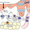
Lupus erythematosus (LE) is a chronic autoimmune disease that has a highly variable range of clinical presentation and course. CLE solely manifests dermatological symptoms, whereas systemic LE (SLE) is a life-threatening disease that commonly affects any organ system, including the skin, joints, and cardiovascular and central nervous systems. Except nonspecific cutaneous manifestations of LE, typical CLE lesions are classified into three subtypes, including acute, subacute, and chronic CLE, that share common histological features characteristic of this disease. The most typical clinical manifestation of acute CLE is a butterfly rash occurring over the bridge of the nose. Subacute CLE represents annular erythema or psoriasis-like scaling erythematous plaques. In chronic CLE, discoid LE (DLE) is the most frequent type and chilblain lupus is its variant. The most prominent histological features of CLE are keratinocyte death and variable inflammation around the dermoepidermal junction. The initial trigger leading to the development of CLE has been believed to be apoptosis of epidermal keratinocytes especially caused by ultraviolet radiation (UVR). This notion is supported by the clinical observation that CLE lesions are preferentially distributed in sun-exposed areas such as face, nose, ears, and neck.
The mechanism underlying the developmental process of CLE has been well studied using experimental photoprovocation. In healthy individuals, the increase in apoptotic keratinocytes is observed only 24 h after UVR exposure and unviable cells completely disappear within 72 h [37], indicating that appropriate phagocytosis efficiently removes apoptotic cells and prevents the release of pro-inflammatory mediators from them. In contrast, apoptotic keratinocytes accumulate up to 72 h after UVR exposure in lupus patients as a result of clearance deficiency [37], which is an intrinsic defect of this disease [46]. When clearance fails, apoptotic cells enter the stage of secondary necrosis. The release of intracellular danger signals from the dying cells potentially plays a crucial role in the breaking of immune tolerance in CLE, but the danger signals from secondary necrotic cells still remain elusive because apoptosis consumes most of well-known danger signals derived from primary necrotic cells. Recent studies have identified danger signals from secondary necrotic cells, including high mobility group Box 1 (HMGB1) associated nucleosomes [69], caspase-cleaved or granzyme B-cleaved autoantigens [54], and uric acid forming monosodium urate crystals in the extracellular space [36]. The detailed mechanism by which these danger signals activate immune cells is largely unknown due to the little information on their cognate receptors, but they activate NFκB and inflammasome in immune cells [12], resulting in the recruitment of monocytes, macrophages, neutrophils, and DCs; production of pro-inflammatory cytokines and chemokines; and upregulation of costimulatory molecules.
Ro52 plays a key part in a series of the pathological events following the initial injuries of epidermal keratinocytes. In CLE lesions, the expression levels of Ro52 are markedly elevated in the cytoplasm of injured keratinocytes, including apoptotic cells and secondary necrotic cells, regardless of the triggers inducing keratinocyte injury [49]. Ro52 is an IFN-inducible E3 ubiquitin ligase and regulates the activity and stability of IFN regulatory factors (IRF) 3, 5, 7, and 8 via ubiquitination and subsequent degradation [13, 15, 24, 25, 35]. Upregulated expression of Ro52 promotes the ubiquitination of these IRFs and suppresses the IFN-dependent signaling pathways. On the other hand, Ro52 promotes apoptosis as shown in a lymphoma-derived B-cell line overexpressing Ro52 [15]. The expression of Ro52 is induced in epidermal keratinocytes injured by UVR exposure within 24 h [49], leading to the suppression of IFN-dependent signaling pathways and the promotion of apoptosis. Because Ro52 binds to the Fc part of any IgG with unexpectedly high affinity comparable to that of the bacterial superantigen protein A [53], Ro52 derived from dying keratinocytes forms ICs with IgG. Furthermore, anti-Ro52 antibody, which is frequently detected in lupus patients, recognizes the Ro52 antigen, resulting in the formation of massive ICs as detected at the dermoepidermal junction by the lupus band test.
ICs containing nuclear constituents might be recognized by pDCs and amplify type I IFN secretion, especially IFN-α [67]. IFN-α generally induces the expression of Ro52 in immune cells [15, 65] and suppresses the ongoing inflammation by inhibiting their proliferation and promoting their apoptosis. Supporting this notion, Ro52 is highly expressed in infiltrating immune cells of psoriasis, lichen planus, and atopic dermatitis as well as CLE [13, 49], and Ro52-deficient mice show uncontrolled inflammation in response to minor skin injury leading to an LE-like condition [13]. In CLE lesions, Ro52 is expressed at high levels particularly in CD4+ T cells, CD8+ T cells, and macrophages [15, 70]. The presence of anti-Ro52 antibody, which neutralizes E3 ubiquitin ligase activity of Ro52 [14], and a genetic polymorphism of Ro52 [17] potentially leading to its altered expression and/or function may explain a part of the mechanism underlying the dysregulated inflammatory process of CLE.
Pro-inflammatory mediators derived from injured and dying keratinocytes activate macrophages and DCs and promote phagocytosis of cell debris and ICs including autoantigens . DCs may present autoantigens to naïve, potentially self-reactive, T and B cells in the lymph node. An attack of the adaptive immune system via autoreactive antibody and effector T cells induces keratinocyte injury and amplifies inflammation. Upregulated expression of CXCL9, 10, 11, and 12 indicates the contribution of Th1 immune response to the development of CLE lesions [16, 40, 44, 70]. In addition to Ro52, genetic polymorphisms are also reported in IRF5 [20, 29] and IRF7 [18]. Therefore, the impairment of IFN-dependent signaling pathways due to altered expression and/or function of Ro52, IRF5, and IRF7 may be a possible predisposing factor for LE.
Based on the available data described above, the most probable trigger for the onset of LE may be the interplay between environmental factors inducing apoptotic keratinocytes, such as UVR exposure, and intrinsic factors, such as clearance deficiency and altered expression and/or function of multiple genes involved in the regulation of the innate and adaptive immune responses.
31.4 Systemic Sclerosis
Systemic sclerosis (SSc) is a multisystem connective tissue disease characterized by immune abnormalities, vasculopathy, and resultant fibrosis of skin and certain internal organs. Although the pathogenesis of SSc still remains unknown, genomewide association studies have implicated polymorphisms in the HLA locus and other immune-associated genes as risk factors for the development of SSc [1, 52, 73], suggesting that the immune system is a central mediator of this disease. Consistently, autoantibody production usually precedes the earliest dermal pathological changes, such as alterations in endothelial cell (EC) function and ultrastructure of the microvasculature [50, 63]. Following and/or in parallel with these vascular changes, inflammatory mononuclear cells (MNCs) infiltrate into perivascular regions [31] and further promote EC damage, leading to the development of vasculopathy characteristic of SSc, such as reduction in the number of capillaries, thickening of arteriolar walls, and intimal fibrosis of small arteries. In parallel with the progression of vasculopathy, dermal fibroblasts are activated especially in the perivascular areas [60]. Further activation of dermal fibroblasts due to their intrinsic abnormalities, such as autocrine TGF-β signaling (described below), and tissue hypoxia eventually results in the establishment and maintenance of extensive skin sclerosis. These sequential pathological events of SSc suggest that the microvascular endothelium is one of the major targets of the immune reaction and a potential trigger for subsequent inflammatory changes and the development of fibrosis.
Although the detailed mechanism causing the initial EC damage remains largely elusive, several lines of evidence suggest the contribution of γδT cells and antiendothelial cell antibodies (AECAs) to this pathological process. In the early edematous phase of the disease, the perivascular infiltrate consists primarily of CD3+ T cells, with CD4+ T cells predominating over CD8+ T cells [43]. Note that there are a number of γδT cells infiltrating in the perivascular areas as well. The majority of γδT cells in the skin as well as in peripheral circulation and bronchoalveolar lavage fluid express the Vδ1 chain in SSc patients, whereas the majority of circulating γδT cells are Vδ2+ in healthy subjects [19]. Furthermore, almost all of the peripheral Vδ1+ γδT cells in SSc patients are positive for the activation marker CD49d [19], which mediates adherence of these cells to ECs by interacting with vascular cell adhesion molecule-1. Considering that circulating γδT cells from early diffuse cutaneous SSc (dcSSc) patients favorably bind to ECs and exert a great cytotoxicity against them [30], γδT cells potentially contribute to an immune-mediated damage to the endothelium in the early pathological process of SSc. Another potential mediator of EC damage is a subset of autoantibodies against heterogeneous antigens on ECs, that is, AECAs, which are detectable in serum of 44–84 % of SSc patients [26, 55, 56]. AECAs induce apoptosis of human dermal microvascular ECs through antibody-dependent NK cell cytotoxicity via the Fas pathway, but not via the perforin/granzyme pathway [62]. Because the cytotoxic effect of AECAs is not observed against human umbilical vein endothelial cells [62], AECAs-dependent vascular injury may at least partially explain the reason why EC damage in early SSc selectively occurs in microvasculature.
EC damage generally induces the expression of cell adhesion molecules and chemokines and promotes the infiltration of inflammatory cells to the perivascular areas. Of note, the comparison of gene expression profiles between peripheral blood mononuclear cells from SSc patients and those from healthy controls indicate overexpression of a cluster of gene-encoding molecules that target these cells to the endothelium in those from SSc patients [66]. Because serum levels of cell adhesion molecules correlate with the severity of tissue fibrosis and/or vascular complications to a variable degree [27, 28, 39, 61, 64], the cell adhesion molecule-mediated interaction between T cells and ECs/other target cells may play a vital role in the development of vascular and fibrotic involvement of SSc.
Tissue fibrosis is the most prominent clinical feature of SSc, which is achieved by fibroblast activation following the initial vascular and immunological events. In the acute inflammatory stage of the disease, fibroblasts expressing type I and type III procollagen are predominantly located around small blood vessels with surrounding infiltration of MNCs [60], suggesting that these immune cells are activated through their interaction with the injured endothelium and subsequently stimulate fibroblasts and enhance their collagen production. In this disease stage, a majority of infiltrating T cells, including Vδ1+ γδT cells, express activation markers, including the early activation marker CD69 [31]. Inasmuch as CD69 regulates cell contact interaction between T cells and other cells, including fibroblasts , activated T cells may affect the activation status of SSc dermal fibroblasts. Notably, the response to activated T cells in a coculture system is different between normal and SSc dermal fibroblasts. In normal dermal fibroblasts, collagen production is suppressed by Th1-polarized cells through membrane-associated IFN-γ [11] and by Th2-polarized cells through membrane-associated TNF-α [10], which overcome a pro-fibrotic effect of IL-4. In contrast, increased collagen synthesis of SSc fibroblasts is more resistant to Th1-polarized cell-mediated suppression and completely unresponsive to Th2-polarized cells [10, 11]. Given Th2 immune polarization during the early active stage of dcSSc (as described below), unresponsiveness to Th2-polarized cell-mediated suppression may contribute to fibroblast activation in early stage of dcSSc. As well as direct interaction, cytokines secreted by activated T cells are involved in the regulation of ECM deposition by fibroblasts [9]. It has been suggested that Th1 cytokines , including IFN-γ, generally decrease ECM deposition, whereas Th2 cytokines , including IL-4, IL-6, IL-10, and IL-13, increase it [9]. A series of studies suggests that the Th1/Th2 paradigm largely contributes to tissue fibrosis of SSc. In the early stage of dcSSc with progressive skin sclerosis, serum levels of IL-6 and IL-10 are significantly elevated, and their levels are decreased to normal levels in the late stage of dcSSc with the improvement of skin sclerosis [58]. Another Th2 cytokine, IL-4, keeps normal levels in the early stage of dcSSc, but is decreased as well in the late stage of dcSSc. In contrast, serum levels of IL-12, a key Th1 cytokine, are decreased in the early stage of dcSSc, then gradually increase in parallel with disease duration and finally reach significantly higher levels than normal controls in the late stage of dcSSc with the resolution of skin sclerosis [42]. Of note, the levels of maximal serum IL-12 levels throughout the disease course inversely correlate with mortality in early dcSSc with diffuse cutaneous involvements. Thus, immune polarization in SSc generally shifts from Th2 to Th1 in parallel with disease duration, whereas the sustained Th2 immune polarization closely associates with exacerbation of the disease. As for Th17 cytokines , the expression levels of IL-17A, but not IL-17F, are increased in the skin of early dcSSc. Furthermore, IL-17A suppresses collagen production in normal dermal fibroblasts, but not in SSc dermal fibroblasts at least partly due to the loss of IL-17 receptor type A [47]. Collectively, Th1/2/17 paradigm regulates tissue fibrosis in SSc.
TGF-β is a key growth factor regulating the activation status of dermal fibroblasts in SSc patients. Although the expression pattern of TGF-β in the lesional skin of SSc is still controversial, TGF-β expression levels generally seem to be higher in patients with early active disease, but weak or undetectable in patients with sclerotic disease. A series of studies regarding the expression profiles of the three isoforms of TGF-β have elucidated the following findings: (i) expression of TGF-β1 and TGF-β2 is most prominent around dermal vessels and is associated with infiltrating MNCs; (ii) staining for all three isoforms of TGF-β is detected in the ECM; (iii) TGF-β mRNA or protein is detected in fibroblasts, particularly those located closely to infiltrating MNCs; and (iv) TGF-β2 transcripts colocalize with α1(I) collagen mRNA and are found in perivascular distribution and surrounded by infiltrating MNCs [22, 38, 51]. Given that the nature of TGF-β is determined by the state of activation and differentiation of the target cells and the presence and concentration of other cytokines and growth factors, TGF-β potentially promotes inflammation by recruiting leukocytes through regulation of cell adhesion molecule expression and creation of a chemokine gradient, by activating leukocytes, and by inducing various pro-inflammatory cytokines and other mediators in an early stage of the disease. In the sclerotic stage, by contrast, SSc dermal fibroblasts are constitutively activated with the pro-fibrotic phenotype quite similar to that of normal fibroblasts treated with TGF-β1 even though the expression of TGF-β is weak or undetectable in the skin [5]. This observation suggests that once activated as a result of vascular and immunological events SSc fibroblasts establish a self-activation system at least partially via autocrine TGF-β signaling . The increased expression of latent TGF-β receptors , including integrin αVβ3, αVβ5, and thrombospondin-1, contribute to this process in SSc dermal fibroblasts [2–4, 6, 45]. These receptors recruit and activate latent TGF-β on the cell surface and efficiently increase the concentration of active TGF-β around SSc fibroblasts. Therefore, dermal fibroblasts may be constitutively activated by autocrine TGF-β in SSc lesional skin.
Another subset of infiltrating immune cells in the lesional skin of early SSc is B cells and plasma cells , which are occasionally, but not frequently seen [50]. Note that a cluster of genes associated with B cells is strongly expressed in lesional and nonlesional skin of SSc patients [71]. Taken together with the evidence that hypergammaglobulinemia, autoantibody production, and CD19 overexpression in B cells are seen in SSc [57], B cells may play a critical part in the pathogenesis of SSc.
Autoantibodies in SSc are classified into two groups: autoantibodies directed against nuclear antigens (topoisomerase I, centromere, RNA polymerase I/III, etc.), and autoantibodies with putative pathogenic roles. Of note, antinuclear antibodies specific to SSc, such as antitopoisomerase I antibody and antinucleolar antibodies , react with nuclear antigens derived from apoptotic cells (e.g., apoptosis of ECs by AECAs) and these ICs induce the production of IFN-α from circulating pDCs [32], which potentially induces autoimmunity in several ways (as already described). The other group of autoantibodies with putative pathogenic roles includes antibodies against fibrillin-1, MMP-1, MMP-3, and PDGR receptor. Antifibrillin-1 antibodies are detectable in more than 50 % of SSc patients and can activate fibroblasts and stimulate release of TGF-β [74]. Antibodies to MMP-1 and MMP-3 may also occur in a high proportion of patients, preventing the degradation of excessive collagen [48, 59]. A putative pathogenic autoantibody to PDGF receptor has been recognized in SSc patients, and implicated in collagen gene overexpression by fibroblasts [7]. Therefore altered B-cell function may be a key link between autoimmunity and tissue fibrosis.
Thus, accumulating evidence suggests the central role of the immune system in the development of vasculopathy and tissue fibrosis in SSc. However, this notion is still largely based on several hypotheses. At this moment, the lack of animal models recapitulating all three cardinal features, including immune abnormalities, vasculopathy, and fibrosis, has hindered further progress in understanding the pathogenesis of this disease.
31.5 Dermatomyositis
DM is an idiopathic inflammatory myopathy characterized by proximal muscle weakness, muscle inflammation, and inflammatory skin rash. DM skin lesions include Gottron’s papules (violaceous papules overlying the dorsal interphalangeal, metacarpophalangeal, elbow, or knee joints), violaceous poikiloderma (atrophic, hypo/hyperpigmented macules with telangiectasia around the anterior neck, chest, shoulders, back, and buttocks), a periorbital heliotrope rash, and periungual telangiectasias. Histopathology of DM skin lesions features an inflammatory infiltrate of CD4+ and CD8+ T cells, pDCs, and neutrophils, along with basal keratinocyte vacuolar alteration with apoptotic keratinocytes, endothelial cell damage, and mucin deposition.
Related posts:


Stay updated, free articles. Join our Telegram channel

Full access? Get Clinical Tree


