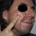
Fig. 1
Patient 60 days after TCA 35% associate to abrasion to treat acne scars
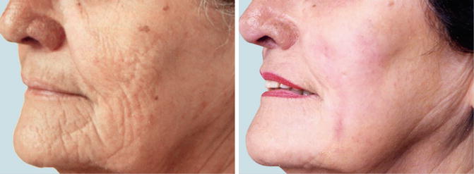
Fig. 2
Patient 60 days after TCA 35% associate to abrasion to treat wrinkles and laxity
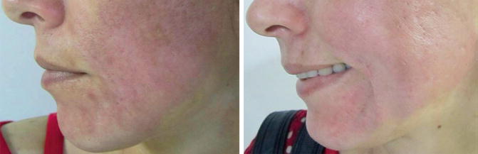
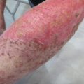
Fig. 3
Patient 30 days and 60 days after procedure presenting dyscromias and persistent erythema
Fundamentals of PCIM
Orentreich and Orentreith (Orentreich and Orentreich 1995) coined the term subcision to describe the subcutaneous incisionless surgery using hypodermic needles for treating depressed scars and wrinkles, aiming to stimulating collagen production. Based on the same principle of rupturing and removing damaged subepidermic collagen and subsequently replacing it with new collagen and elastin fibers, other authors confirmed this initial study. More recently, a system of microneedles applied to the skin was proposed, with the objective of generating multiple micropunctures, long enough to reach the dermis and cause bleeding, triggering inflammatory response that induces collagen production (Camirand and Doucet 1997; Fernandes 2006).
The percutaneous collagen induction (PCI), as the technique has been called, begins with loss of the cutaneous barrier integrity (causing keratinocyte dissociation), resulting in release of cytokines such as interleukin-1α (predominantly), interleukin-8, interleukin-6, TNF-α, and GM-CSF and leading to dermal vasodilation and migration of keratinocytes, a process that restores the epidermal damage (Bal et al. 2008). For didactic purposes, three stages of the healing process following trauma with needles can be clearly delineated. The first stage (injury stage) is characterized by the release of platelets and neutrophils (which are responsible for releasing growth factors that act on keratinocytes and fibroblasts, such as transforming growth factors α and β (TGF-α and TGF-β), platelet-derived growth factor (PDGF), protein III (activator of connective tissue), and connective tissue growth factor). In the second stage (healing stage), neutrophils are replaced by monocytes, and angiogenesis, epithelialization, and fibroblast proliferation take place, followed by the production of type III collagen, elastin, glycosaminoglycans, and proteoglycans. Concomitantly, fibroblast growth factor, TGF-α, and TGF-β are secreted by monocytes. Roughly 5 days after the injury inflicted, the fibronectin matrix is completely formed, allowing the deposition of collagen directly beneath the basal layer of the epidermis. In the third stage (maturation stage), type III collagen, which is prevalent in the early phase of the healing process, is slowly replaced by type I collagen (which lasts longer and persists for a period ranging from 5 to 7 years) (Fernandes and Massimo 2008; Aust 2008a, b).
In order for this inflammatory sequence of events to take place, the trauma caused by the needle must reach a depth of 1–3 mm, and the epidermis must be preserved (only perforated and not removed). Hundreds of microlesions are created, resulting in columns of blood collected in the dermis, accompanied by edema of the treated area and virtually immediate hemostasis. The intensity of these reactions is proportional to the length of the needle used in the procedure. For instance, a 1 mm depth entails an almost microscopic hematoma, while that resulting from a 3 mm depth can be seen with the naked eye and can persist for hours. Nonetheless, it is necessary to understand that the needle does not penetrate completely during the rolling process. It is estimated that a 3 mm long needle penetrates only 1.5–2 mm (or roughly 50–70% of its total length). Therefore, with a 1 mm long needle, the injury caused to the skin would be limited to the superficial dermis, resulting in a more limited inflammatory response than that caused by a longer needle (Aust 2008b; Fabroccini and Fardella 2009; Lima et al. 2013; Vasconcelos et al. 2013; Lv et al. 2006; Vandervoort and Ludwig 2008).
Characteristics of PCIM
The device used to perform PCIM comprises a polyethylene roll studded with sterile stainless steel needles symmetrically aligned in rows, totalizing around 190 units (a number that may vary depending on the manufacturer). The length of the needles is fixed throughout the structure of the roll and varies from 0.25 to 2.5 mm, according to the model. The procedure is usually well tolerated under local anesthesia, with needles not exceeding 1 mm in length (Fig. 4). For greater lengths, anesthetic blockade supplemented by infiltrative anesthesia is recommended (Fernandes 2006).
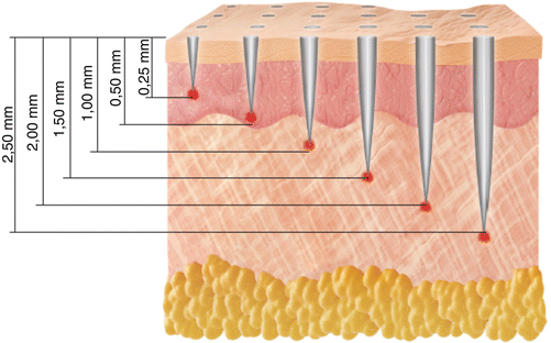
Fig. 4
Correlation between the length of needles and penetration into skin
Aiming to provide more comfort for the patient in situations of prolonged surgical time and deeper injury, local anesthesia with sedation is recommended. PCIM is a technique-dependent procedure, and the final outcome is directly influenced by familiarization with the device used and mastery of the recommended technique. The vertical pressure exerted on the roller must not be strong to avoid damaging deeper anatomical structures and excessive pain. The device is recommended to be positioned between the thumb and index finger—as if holding a hashi—controlling the force with the thumb. The back and forth movements must imprint a uniform pattern of perforations (resembling petechiae) throughout the treated area. In order to achieve this, 10–15 passes in the same direction must be made, and at least four crossing passes in the rolling areas seem to be sufficient. In theory, 15 passes allow a controlled damage that corresponds to 250–300 punctures/cm2.
The time that the petechiae pattern takes to arise varies according to the thickness of the treated skin and the selected needle’s length. Therefore, a thinner and looser skin, which is usually photodamaged, will present a uniform petechiae pattern earlier than a thicker and fibrotic skin, which is commonly observed in patients with acne scars, for example. In this manner, the choice of the needle’s length depends on the type of the skin to be treated and the ultimate goal of the procedure. There is not yet a classification correlating the length of the device’s needles to the depth of the expected damage in the treatment (Fernandes 2006; Fernandes and Massimo 2008).
Related posts:


Stay updated, free articles. Join our Telegram channel

Full access? Get Clinical Tree


