Chapter 24. Closed Approach to Rhinoplasty
Rodger H. Brown, MD; Daniel A. Hatef, MD; Jamal M. Bullocks, MD; Samuel Stal, MD
INDICATIONS
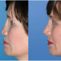
The debate about whether to use the open or endonasal approach to rhinoplasty has been ongoing on for many years. The majority of rhinoplasty procedures can be performed in an endonasal or closed approach, but some surgeons believe there are advantages to using the open approach in more difficult cases such as secondary or cleft rhinoplasties. The open approach involves an external incision across the columella that will lead to a scar. This incision usually heals well, but it can occasionally lead to an unsightly scar and/or notching (Fig. 24-1).
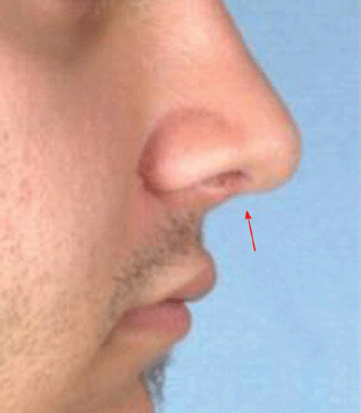
Figure 24-1 Picture of notching after open rhinoplasty incision across the columella.
The patient’s nasal deformity should be assessed preoperatively, and the surgeon needs to determine whether he or she is capable of correcting that deformity through an endonasal approach or whether wider exposure will be needed. Most experts would agree that simple dorsum, tip, and septal problems can easily be addressed with the closed approach. In more complex problems, the decision between closed versus open depends on the extent of the deformity, the techniques planned, the patient’s desires, and the surgeon’s experience.
PREOPERATIVE PREPARATION
The preoperative consultation should consist of a complete examination of the external nasal features, the internal nasal anatomy, and the relationship of the nose to other facial features. In endonasal rhinoplasty, it is important to be able to look at the nose externally and understand what underlying structural abnormality is causing each deformity. Unlike in open rhinoplasty, there is limited ability to visualize the underlying framework during the operation. Correct evaluation and diagnosis of the underlying deformity allows the surgeon to plan what procedures and techniques will be used during the operation. If the preoperative evaluation is incorrect, the procedures performed will not correct the deformities and may make them worse. This is one of the biggest criticisms of the closed approach, but if one becomes skilled at the preoperative evaluation and diagnosis, the endonasal approach can allow for a shorter and less-traumatic approach to rhinoplasty, yielding equal or superior results.
The preoperative exam should be performed in the same organized manner with each patient to develop a routine. This will make the surgeon more efficient and accurate in assessment and diagnosis. One good way to approach it is in a top-down, external-to-internal fashion. Begin with evaluation of the radix, followed by the dorsum, tip, columella, ala, nostrils, and external nasal valve. Examine the internal structures, which include the septum, turbinates, and the internal nasal valve, with a nasal speculum and adequate lighting.
ANESTHESIA
Endonasal rhinoplasty may be performed under local anesthesia only, local anesthesia and sedation, or general anesthesia. Regardless of technique, the infiltration of local anesthetic with epinephrine is effective to limit bleeding, allow better visualization, and better define tissue planes.
The local anesthetic preparation is begun by packing the nose with cottonoids soaked in 4% cocaine or Afrin (oxymetazoline) to create a vasoconstricted state. Afrin causes vasoconstriction only, while cocaine will cause a more intense vasoconstriction and also act as a local anesthetic. Cocaine has the potential for causing cardiac complications, and should be used with caution in patients at risk for such complications; the dose should be strictly limited to 4 mg/kg. Next, the nasal structures are infiltrated with 1% lidocaine with 1:200,000 epinephrine. Usually only 5 to 10 mL are needed. Injection of too much local anesthetic can distort the soft tissues and make intraoperative evaluation of results difficult. The local anesthetic solution should be injected at the radix, along the dorsum, along the ascending processes of the maxilla at the proposed osteotomy sites, along the septum, the alar-facial grooves, and a small amount over the lower lateral cartilages and tip. Small amounts should also be injected at the sites of any other proposed incisions.
POSITION AND MARKINGS
The patient should be placed in the supine position with a shoulder roll to create slight extension of the neck. The entire face should be prepped in the usual sterile fashion. If the patient is under general anesthesia, the eyes should be protected with lubricant and an occlusive dressing or corneal protectors. A sterile adhesive drape can be used to further isolate the mouth and endotracheal tube from the operative field. Finally, sterile towels are used to create a head drape as in other facial surgery. Given that all incisions are endonasal in the closed approach, no markings are absolutely necessary, but some find it helpful to mark the borders of the lower lateral cartilage, the midline of the tip, and the caudal end of the nasal bones.
DETAILS OF PROCEDURE
Incisions
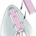
An infra-, intra-, and/or intercartilaginous incision is made using a no. 15 blade with a sharp double hook to help evert the alar rim for exposure. The infracartilaginous, or marginal, incision is placed at the caudal border of the lower lateral cartilage (Fig. 24-2
Related posts:


Stay updated, free articles. Join our Telegram channel

Full access? Get Clinical Tree


