(1)
Department of Dermatology and Allergology Biederstein, Technische Universitaet Muenchen (TUM), Munich, Bavaria, Germany
(2)
Christine Kuehne Center for Allergy Research and Education (CK-CARE), Hochgebirgsklinik (High Altitude Hospital), Davos, Switzerland
Describing the clinical symptomatology of this skin disease is more difficult than in other dermatoses. Maybe this is why the big masters of morphology in dermatology preferred other skin diseases like psoriasis, bullous diseases, or lichen planus with clear-cut primary lesions and well-described secondary skin changes.
Atopic dermatitis often represents as a “diffuse” skin condition. The definition of the whole disease seems to be as unprecise as the borders of affected skin areas. Sometimes the term “involved” skin is questionable, especially when the dermatohistopathological investigations used show signs of inflammation also in clinically “uninvolved” skin areas (Kunz and Ring 2000).
The susceptibility for environmental influences, especially on the psychosocial level, underlines the strong variability of this dermatosis. Itch crisis can elicit attacks of rage. Rage can elicit attacks of itch and scratching. In addition, this disease manifests itself differently in different age groups and different body regions.
For logic didactic reasons, this chapter will describe the clinical symptomatology of atopic dermatitis in the following order:
Actual symptoms of disease
Minimal manifestations
Stigmata of atopic constitution
Possible complications
However, it is clear that these four phases often exist simultaneously and are variable over time even with regard to stigmata.
2.1 Actual Symptoms of Disease
2.1.1 Primary Lesion “Itch”
Normally dermatologists in the description of a skin disease start with the “primary lesion.” There is no consensus with regard to the primary lesion of atopic eczema; it is described as erythema, papule, and seropapule. Some authors, starting with Jacquet (1904) and the French School, stress the symptom “pruritus.” According to good tradition the author says, together with Saint John the Evangelist or Johann Wolfgang von Goethe (Faust): “In the beginning there was the itch!” (Rajka 1980, 1989; Ring 1982a).
Rarely it is possible to observe the acute development of an eczematous skin lesion in atopic dermatitis directly; however, sometimes—e.g., with your own children or under provocation tests in the hospital—it is possible. From this experience it can be concluded that human beings, who never before have shown any signs of eczematous skin changes, triggered by some environmental or psychologic factors suddenly develop itch and start scratching on apparently normal skin. Only hours later the typical clinical manifestations of atopic dermatitis develop. These phenomena are especially common in the areas mostly affected such as the elbow, knee flexures, or eyelids. It is a pity that there is little scientific documentation with regard to these early manifestations in the individual initial phase. Many colleagues, parents, and patients have made this experience.
Itch as “dermatose invisible” is the primary lesion of atopic dermatitis which, via the scratch reaction, then gives rise to the characteristic morphologic skin changes (Fig. 2.1). Besides, it is possible that primarily skin changes occur which go along with increased itch. The borders are fluent. A typical atopic eczema without itch is extremely rare, if not inexistent. Since the French School at the beginning of the twentieth century, the central importance of itch for atopic dermatitis has been stressed by many authors (Borelli and Schnyder 1962; Braun-Falco and Ring 1984; Breit 1997; Engman et al. 1936; Haxthausen 1957; Kimura and Miyazawa 1989; Koblenzer 1999; Rajka 1989; Ring et al. 2006; Talbot 1918).


Fig. 2.1
(a) Itch sensation painted by a 9-year-old girl suffering from atopic dermatitis. (b) The primary lesion “itch” leads via scratch effects to the typical skin changes of atopic dermatitis
Itch is the most agonizing symptom of manifest disease and often is perceived as “incurable.” It leads to sleep loss, nightly scratch attacks with bloody bedding, a feeling of “powerlessness,” because it cannot be stopped voluntarily. Unfortunately this symptom is not yet taken seriously by many physicians and the society, contrary to the other main subjective symptom “pain.” Regarding the latter, everybody is full of compassion (Ständer et al. 2010). I personally know patients who wanted to commit suicide because of severe itch, a fact also observed by other authors (Dieris-Hirche et al. 2009).
The old sentence “It is not the rash which itches, but the itch that rashes” (Engman et al. 1936) illustrates the situation. Early attempts of treatment 80 years ago quite brutally used procedures like fixation of children in bed or applying plaster to their extremities, so that scratching became impossible. Lesions in unreachable skin areas were indeed healing faster. Pathophysiologically this indicates that by scratching proinflammatory substances may be released (see Chap. 3).
Today the treatment of itch is the focus in the management of atopic dermatitis, and the prevention of direct scratch effects by mechanical procedures such as wet wraps with tube bandages plays a central role (see Chap. 5).
2.1.2 Infiltrated Erythema
Mild cases or the beginning of an exacerbation become manifest in flat, mildly infiltrated, erythematous patches with only little epidermal involvement which in the course can be excoriated and change to oozing crusting areas (Fig. 2.2).


Fig. 2.2
Infiltrated erythema. (a) On the trunk in an infant. (b) In the elbow in a 1-year-old child
In the severity scoring of the SCORAD (see Sect. 2.8.2, page 64), these criteria “erythema,” “edema and papule,” but also “lichenification” are enlisted.
It is important to stress that there are these manifestations since they are not clearly mentioned in many textbooks.
2.1.3 Erosive, Excoriated Erythema
Especially in childhood, often patchy or punctate erosive or excoriated erythematous skin changes with mild scaling are observed, going along with intense itch (Fig. 2.3).
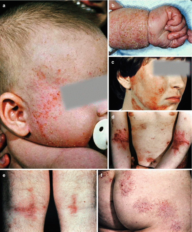
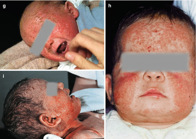


Fig. 2.3
Erosive excoriated eczematous skin changes in atopic dermatitis. (a) Facial eczema in an infant. (b) Scaling hand and forearm eczema in an infant. (c) Diffuse erythematous excoriations in the face. (d) Marked erythematous flexural eczema in the elbow. (e) Punctate erosive and excoriated skin changes in the knee joints. (f) Diffuse, partly nummular appearing erythemata with punctate erosions on the buttocks. (g) Massive facial eczema in an infant. (h) Facial eczema in an infant. (i) Facial eczema in a small child
Histologically these lesions are characterized by spongiotic changes in the epidermis. This form often has been called “eczematoid type” in the restricted sense. This may have contributed to dissatisfaction with the name “eczema” for the whole disease and may be a reason for the terminology and preference of the unprecise term “dermatitis.”
In the broader definition of eczema (see Sect. 2.8.2), it becomes clear that in the local and timely course, the polymorphy of symptoms in eczema also includes other morphologies like lichenification, prurigo-type excoriated papules, etc.
Maybe strongly oozing and crusty skin changes in childhood represent signs of superinfection with, e.g., Staphylococcus aureus (see Sect. 2.6).
This morphology also can be observed in circumscribed skin lesions with nummular variants (see Page 37). Whether nummular eczema in childhood represents an own identity is still to be discussed. In the opinion of the author, this nummular variant is a subtype of atopic eczema in childhood.
2.1.4 Lichenification
The more chronic the skin changes, the more it shows a tendency to lichenification. This means a coarsening of the skin signs with thickening of the skin and livid red color “purple.” These skin changes occur predominantly in the flexures of the big joints (elbow, knee flexure), but also as large plaques on the dorsum of the hands and feet. They also represent most likely the consequence of long-lasting scratch effects (Fig. 2.4).
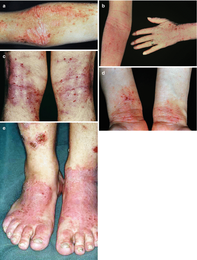

Fig. 2.4
Lichenification in chronic atopic dermatitis. (a) Lichenified flexural eczema in the elbow. (b) Lichenified eczema on the wrist. (c) Lichenified eczema in the knee joints. (d) Lichenified skin areas on the wrists. (e) Massive lichenification on the dorsum of the feet in an adult patient with atopic dermatitis
The entity of lichen simplex chronicus (Vidal) or “circumscribed neurodermatitis” can be regarded as a subgroup of localized variants of atopic eczema; however, this is a matter of discussion.
Lichenification can be provoked by scratching as could be shown in classic experiments with the use of a scratch machine with a pressure of 75 g on the skin. After 60–90 h lichenification could be provoked (Goldblum and Piper 1954).
2.1.5 Prurigo Type of Atopic Eczema
When the follicular affection in chronic courses is prominent especially in adults, a variant can be observed which shows the clinical symptoms of prurigo; it is relatively marked by strongly itching papules or nodules with a diameter of 0.5–3 mm and central excoriation and crusting. This variant most likely gave rise to the term “prurigo Besnier” and can be found commonly in the severe manifestations in adults (Fig. 2.5).
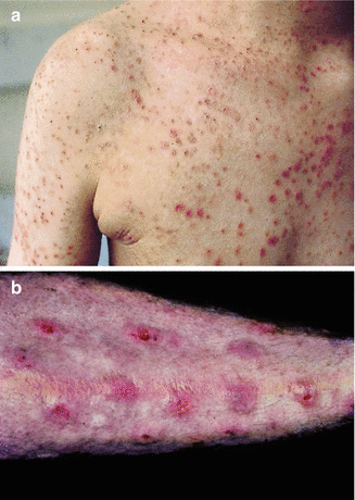

Fig. 2.5
Prurigo type of atopic dermatitis. (a) Centrally excoriated and crusted and papules and nodules in a 17-year-old girl with severe atopic dermatitis. (b) Extremely itchy excoriated papules on the trunk
2.2 Minimal Manifestations
Atopic eczema can change in course and localization and also in morphology over a lifetime. There are certain localizations where so-called minimal manifestations are typical: they can occur alone or sometimes in combination with other skin changes (Herzberg 1973). The affection of very small skin areas can lead to severe impairment of quality of life.
2.2.1 Minimal Manifestations in the Head Area
2.2.1.1 Lid Eczema
Sometimes only the eyelids are affected, especially in cases where aeroallergens play a causal role, sometimes also in food-induced reactions. The more chronic the course, the more difficult the treatment. Already very little contact elicits itch. Through the itch-scratch cycle, a vicious cycle develops with continuous deterioration (Fig. 2.6).
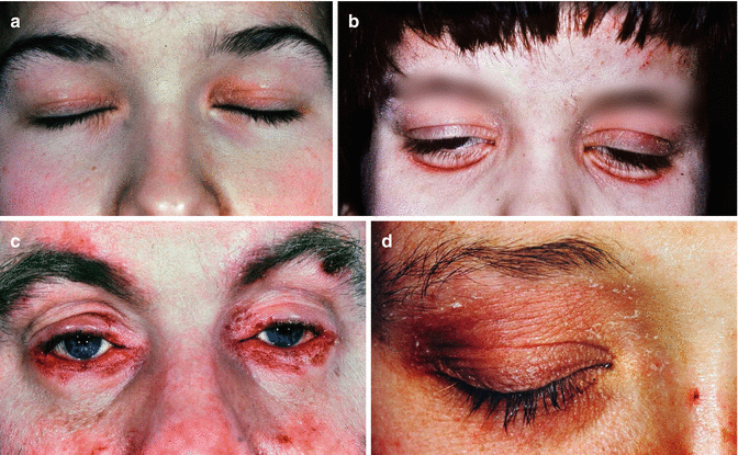

Fig. 2.6
Lid eczema in atopic dermatitis. (a) Upper lid eczema. (b) Lid eczema with edematous swelling. (c) Massive lid eczema of the lower lids. (d) Massive upper lid eczema
2.2.1.2 Cheilitis Sicca and Perlèche
Dry lips—sometimes also with a median fissure of the lower lip—are characteristic signs of atopic eczema (Fig. 2.7), going along with fissures at the lateral angles of the mouth (perlèche) and often superinfection Angulus infectiosus (Candida albicans) (Temime and Oddoze 1970).


Fig. 2.7
Dry lips. (a) Cheilitis sicca with median lower lip fissure. (b) Cheilitis sicca in an adult patient with atopic dermatitis
Sometimes, in the area surrounding the red part of the lip, there is an irritative eczema caused by increased lip licking (lip lick eczema) (Fig. 2.8), in infants sometimes also called suck eczema with a perioral inflammatory reaction.


Fig. 2.8
Lip sucking eczema. (a) Lip sucking eczema with hyperpigmentation. (b) sharply marginated perioral lip eczema and cheilitis sicca
2.2.1.3 Infranasal Erosion
In persons with concomitant respiratory allergy such as rhinoconjunctivitis and chronic nasal secretion or itch, sometimes erythema and fissures on the nasal entrance develop.
2.2.1.4 Infra-auricular Fissures
Infra-auricular fissures are very common at the region where the earlobe starts, which can sometimes be superinfected. It might be interesting how dressing habits—as, e.g., the rapid pulling of a T-shirt over the head of the child—can contribute to these infra-auricular fissures (Fig. 2.9).
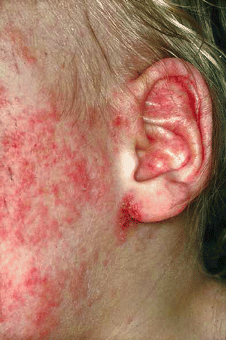

Fig. 2.9
Infra-auricular fissure in atopic dermatitis
2.2.1.5 Retroauricular Intertrigo
Similarly, signs of oozing intertrigo can be found behind the ear (Fig. 2.10).
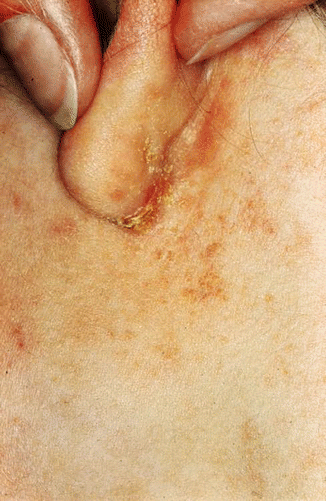

Fig. 2.10
Retroauricular fissure in atopic dermatitis
2.2.1.6 Excoriations in the Scalp
Sometimes extremely itching skin lesions with nodules and crusts and excoriated areas can be found in the scalp as a single manifestation of atopic eczema; severe cases sometimes have been called “neurotic excoriation.”
2.2.2 Minimal Manifestations on the Trunk and Extremities
2.2.2.1 Finger and Toe Eczema
Dry finger and toe changes (pulpitis sicca) can be observed especially in winter and in small children which have been described by Möller as “atopic winter feet” (Möller 1972) and sometimes were misdiagnosed as fungal infection. These changes also can look similar to dyshidrosis lamellosa sicca (Fig. 2.11).
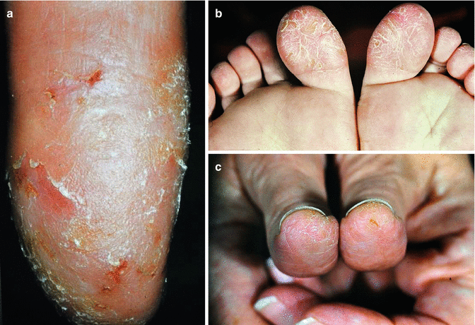

Fig. 2.11
Finger and toe eczema. (a) Dry finger tip eczema. (b) Pulpitis sicca in atopic foot dermatitis (“atopic winter feet”). (c) Painful eczema of the thumbs
2.2.2.2 Involvement of Nails
Strong eczematous changes on the fingers and toes can affect nail growth by matrix involvement with development of horizontal lines (Fig. 2.12a). Sometimes also paronychia develops in atopic eczema with or without superinfection (Fig. 2.12b).


Fig. 2.12
Nail involvement in atopic dermatitis. (a) Eczema nails with strong horizontal grooves representing disturbance of nail growth in the matrix. (b) Paronychia in atopic dermatitis
Atopic dermatitis manifestations also can affect the eponychium, leading to spontaneous loss of eponychium as “epoonychitis sicca” (Ring 2005).
Inspection of nails can reveal much about scratching habits (e.g., subungual crusts or shiny nails when the skin is rubbed) (Fig. 2.13).


Fig. 2.13
Inspection of the nails allows information on the type of scratching. (a) Blood under the nail means heavy excoriation. (b) Shimmering nails can be derived from superficial abrasion
2.2.2.3 Juvenile Plantar Dermatosis
2.2.2.4 Eczema of the Mamilla
Mamillar eczema in atopics is a very chronic and localized variant (Fig. 2.14). It has to be evaluated in differential diagnosis in order to be distinguished from M. Paget or scabies.
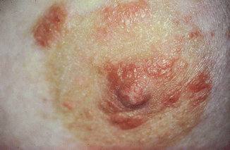

Fig. 2.14
Mamillar eczema in atopic dermatitis (when it is one-sided, scabies or Morbus Paget always have to be considered)
2.2.2.5 Localized Genital Eczema
Similarly there are cases with exclusive affection of the genital regions as vulvar or scrotal eczema with severely lichenified variants in adults and strong psychosomatic involvement (Fig. 2.15).
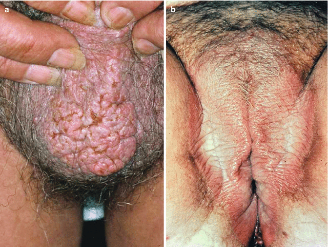

Fig. 2.15
Atopic dermatitis in the genital area. (a) Chronic scrotal eczema. (b) Chronic vulvar eczema
2.2.2.6 Pityriasis Alba
The occurrence of whitish mildly scaling and relatively marked patches especially in light exposed skin areas during summertime is called pityriasis alba and leads to suffering in many patients (Watkins 1961), especially in colored skin (Mazenga, personal communication 2005). Most likely it is due to minimal inflammation with superficial whitish scaling (Fig. 2.16).
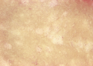

Fig. 2.16
Pityriasis alba in atopic dermatitis
2.2.3 Variants and Special Forms
2.2.3.1 Dermatitis Papulosa Juvenilis
This special variant of atopic eczema goes along with development of fine papular skin changes, sometimes hypopigmented and often at the extensor sides of extremities in childhood (Bork and Hoede 1978; Fölster-Holst et al. 1996). This manifestation can be found under different names (summertime pityriasis of the elbow and knee, dermatite du Tobogan, sandbox dermatitis, frictional lichenoid eruption, recurrent papillary eruption of childhood) (Dupré et al. 1974). Often the skin changes occur predominantly in spring and summer and are associated with pollen exposure. Also mechanic components like friction and sensitivity to UV light have been discussed in the pathophysiology.
2.2.3.2 Follicular Variant of Atopic Dermatitis (Patchy Pityriasiform Lichenoid Eczema)
Kitamura, Takahashi, and Sasagawa described a skin condition in Japan as plaque-type lichenoid squamous dry skin changes in children with follicular involvement and occurring predominantly on the extensor sides, sometimes similar to “goose pimples.” This variant also seems to occur more often in the winter and shows improvement in the summer (Fig. 2.17) (Kitamura et al. 1950; Sasagawa 1967; Wüthrich and Schnyder 1981).
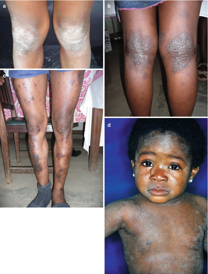

Fig. 2.17
Patchy pityriasiform lichenoid eczema as special variant of atopic dermatitis appearing on the extensor sides of the joints. These skin changes are particularly common in dark skin. (a) Lichenoid papules and plaques on the extensor side of the knee. (b) Atopic dermatitis of the knee joints in an African patient from Tanzania. (c) Atopic dermatitis (nummular pattern) on both legs in an African patient from Tanzania (With friendly permission of P. Schmid-Grendelmeier). (d) Facial eczema in an African child
In the differential diagnosis, follicular dermatoses like lichen nitidus, Gianotti-Crosti syndrome, or follicular hyperkeratosis in certain forms of ichthyosis have to be considered.
2.2.3.3 Nummular Variant of Atopic Eczema
Especially in childhood, nummular eczema can occur with sharply demarcated nummular erythematous skin changes with scaling and excoriation and oozing aspect. Especially the lower extremities, but also the arms and trunk, can be involved. While in adults nummular eczema seems to be an own entity, it is the opinion of the author that nummular eczema in childhood represents a subvariant of atopic dermatitis (Fig. 2.18).
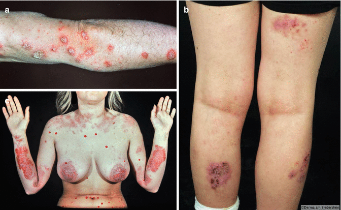

Fig. 2.18
Nummular variant of atopic dermatitis. (a) Scaling nummular lesions in atopic eczema. (b) Nummular eczema of the legs in atopic dermatitis. (c) Sharply marginated massive atopic eczema with nummular distribution
2.2.3.4 Special Considerations in Infants
Atopic eczema most commonly develops at the end of the first trimenon and then occurs in the face and extensor surfaces; commonly the so-called cradle cap is the first manifestation (Fig. 2.19). In the diaper area there are various differential diagnoses to atopic eczema. Interestingly, the diaper area itself is often uninvolved (“diaper sign”) (see Sect. 2.4).
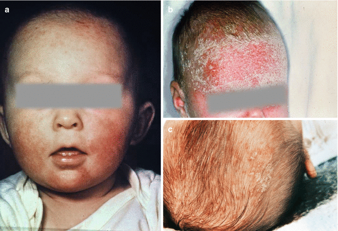

Fig. 2.19
Atopic dermatitis in infants. (a) Facial eczema in infants. (b) Cradle cap (“Crusta lactea” reminding of crusted milk in a pan). (c) Cradle cap on the scalp
2.2.4 Special Localizations
2.2.4.1 Face
In many patients, atopic dermatitis only affects the face (Fig. 2.20).
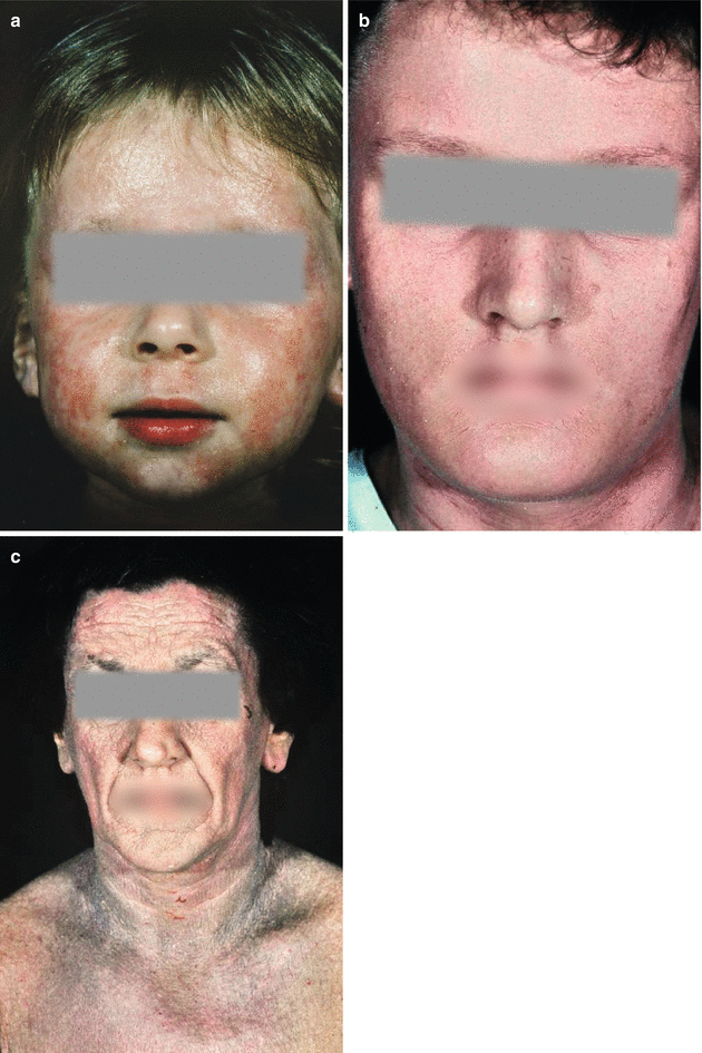

Fig. 2.20
Atopic dermatitis in the face. (a) in childhood. (b) In adulthood. (c) Massive atopic dermatitis of the face with lichenification
2.2.4.2 Neck
Apart from the face, also the neck region is affected in many patients which can be regarded as a “big flexure” (Fig. 2.21). In chronic eczema, the clinical manifestation of a “dirty neck” develops as a postinflammatory hyperpigmentation, sometimes without real symptoms (Colver et al. 1987) (Fig. 2.22).
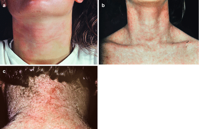
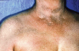

Fig. 2.21
Atopic dermatitis of the neck. (a) Atopic dermatitis of the neck region. (b) Scaling atopic dermatitis of the neck. (c) Atopic dermatitis of the neck with lichenification

Fig. 2.22
“Dirty neck” following postinflammatory hyperpigmentation in atopic dermatitis
2.2.4.3 Hand Eczema
Often the hands and feet show the main manifestation of atopic eczema, with or without concomitant contact allergy. Here scaling eczematous skin changes and dry chronic skin lesions with lichenification, but also, when the palms and soles are involved, dyshidrosis and erosive crusty skin changes can be observed, leading to hyperkeratotic fissuring manifestations (Fig. 2.23). Often the nails are also affected (Fig. 2.24).
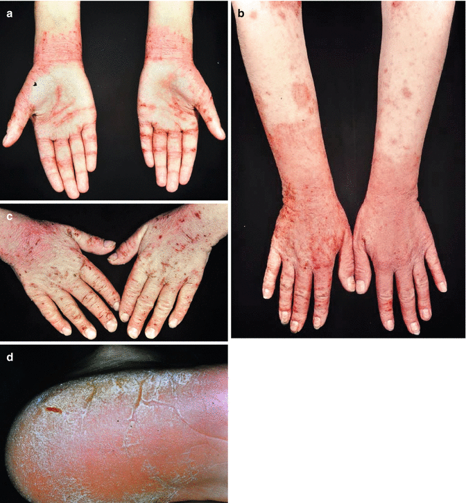


Fig. 2.23
Hand and foot eczema in atopic dermatitis. (a) Hand eczema with palmar involvement. (b) Dorsum of the hands with strong excoriations. (c) Dry atopic dermatitis of the dorsum of the hands. (d) Foot eczema with deep fissures

Fig. 2.24
Nail changes in hand eczema. (a) Eczema nails. (b) Nail dystrophy in eczema
Hand eczema represents a special problem as one of the most common occupational skin diseases and is pathogenetically classified between irritant, contact allergic, and atopic dermatitis. Furthermore, sometimes the differential diagnosis to psoriasis of the palms and soles may be difficult. In a retrospective study, the most frequent subdiagnoses of chronic hand eczema were allergic contact dermatitis, allergic plus irritative contact dermatitis, irritant contact dermatitis, atopic hand eczema, atopic plus irritant hand eczema, vesicular hand eczema (dyshidrotic = pompholyx) and hyperkeratotic eczema (Diepgen et al. 2009).
2.2.4.4 Erythroderma
Atopic dermatitis also can manifest as erythroderma with involvement of the whole integument in the sense of an exfoliative dermatitis with redness and scaling. Erythroderma by definition is a clinical condition which often cannot be attributed to a single disease at first glance. Several diseases can lead to erythroderma:
Allergic contact dermatitis
Psoriasis
Pityriasis rubra pilaris
Cutaneous T-cell lymphoma (Sezary syndrome)
Idiopathic melano-erythrodermia cachectica
Exanthematous drug eruption
2.2.5 Summary
Contrary to other dermatoses, atopic dermatitis does not show a clear-cut morphology. The primary lesion can best be described as itch which then via scratching leads to the formation of skin changes. Several variants with “infiltrated erosive erythema,” “chronic lichenification,” or “prurigo type” of atopic dermatitis can be observed. Exudative eczematous skin changes can be found predominantly during childhood or when superinfection occurs. Minimal manifestations comprise infra-auricular fissures, cheilitis sicca, infranasal erosion, eyelid eczema, finger or toe eczema, and pityriasis alba.
2.3 Stigmata of Atopy
Contrary to actual signs and symptoms of disease and minimal manifestations, the so-called stigmata represent characteristics of the organ skin, not necessarily going along with feeling sick nor being sequels of the disease. Rather they can be regarded as constitutional signs of an “atopic diathesis”; thus, they can give the experienced physician valuable information without additional laboratory diagnostics or history analysis with regard to the existence of atopy in the individual.
Table 2.1 shows the most common stigmata which also have been quantitatively evaluated in epidemiologic studies.
Table 2.1
Stigmata of atopic dermatitis/eczema
Dry skin (sebostasis, xerosis) |
Hyperlinearity of palms and soles (“ichthyosis hands or feet”) |
Linear grooves of fingertips |
Atopy fold of the eyelid (Dennie-Morgan) |
Scattering of lateral eyebrow (Hertoghe) |
Low temporal hairline |
Facial pallor with periorbital halo |
White dermographism |
Delayed blanch reaction after acetylcholine |
2.3.1 Dry Skin (Xerosis)
Dry skin is a major characteristic of atopic dermatitis, also called sebostasis or xerosis; it is visible as rough skin, sometimes slightly scaling (Fig. 2.25). The term “dry” skin can be questioned pathophysiologically since, in investigations with regard to the percentage of water in the skin, no real decrease has been found, but an increased transepidermal water loss (TEWL) due to disturbed skin barrier function. Furthermore, mild signs of inflammation have been found in “dry skin of atopic dermatitis” in uninvolved areas (Uehara 1985).
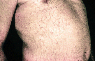

Fig. 2.25
Dry skin, eczema craquelée
The term sebostasis indicates a diminished sebum secretion which is controversial. The lipid content of the epidermis has been investigated by several groups and found to be altered especially with regard to different patterns of ceramides (see Chap. 3).
Finally a major characteristic is roughness which can be recognized by the patient and physician and feels like “dryness.” It is not clear whether this “dry” aspect actually corresponds to the deficiency of the epidermal protein filaggrin (see Chap. 3) found in many patients with atopic eczema.
2.3.2 Ichthyosis Hands and Feet (Hyperlinearity of Palms and Soles)
This stigma is well known in patients with the autosomal dominant disease ichthyosis vulgaris. Marked linear furs or lines can be seen, partly in a bizarre or linear configuration mostly on the palms, but in infants commonly also on the soles (Fig. 2.26).
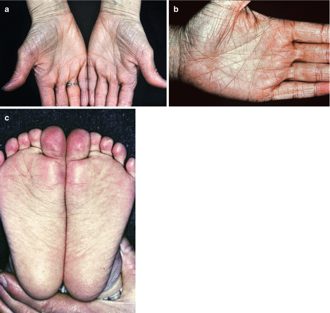

Fig. 2.26
Hyperlinearity of palms and soles (ichthyotsis hands/feet). (a) Massive skin changes in the palms. (b) Hyperlinearity of hand. (c) Ichthyosis feet
This sign, first described by Leutgeb et al. (Leutgeb et al. 1972), was regarded for a long time as proof for concomitantly occurring ichthyosis vulgaris and can now better be explained by the filaggrin deficiency in heterozygous states in many atopic dermatitis patients. Only in a small part of eczema patients the typical signs of ichthyosis vulgaris can be seen in histopathology or by electron microscopy, namely, abnormal keratohyalin granules (Fartasch et al. 1989).
A subgroup of ichthyosis hands or feet can be seen in linear grooves often horizontal to the papillary lines on the fingertips (Fig. 2.27).
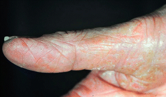

Fig. 2.27
Linear grooves on the finger tips
2.3.3 Infraorbital Fold (Atopy Fold, Dennie-Morgan)
This sign has first been described in a letter by Dr. Morgan as “definite wrinkle just beneath the margin of the lower lid of both eyes,” who mentioned it to Dr. Dennie (Morgan 1948) as a sign of atopy. The fold can be single or double. Mostly it is present on both lower eyelids, sometimes, however, only unilaterally. The exact definition implies a clear-cut double fold beginning mostly at the inner (medial) canthus and reaching laterally at least beyond the center of the pupil. The atopy fold must not be confounded with the physiological sulcus palpebralis inferior (Przybilla et al. 1991; Uehara 1981) (Fig. 2.28). Many authors regard the Dennie-Morgan fold as a sequel of lid eczema due to increased rubbing and scratching. However, clear-cut clinical observations have shown that this stigma can be present many years before the first occurrence of atopic dermatitis, mostly starting in other body areas like the elbows or knee joints and without lid eczema.


Fig. 2.28
Dennie-Morgan fold (atopy fold). (a) Double fold starting on the inner cantus reaching over the middle of the pupil. (b) Atopy fold in a child with atopic dermatitis
The atopy fold is also present in respiratory atopic diseases. In Asian patients, the infraorbital fold seems to be a less suitable marker.
2.3.4 Periorbital Halo and Facial Pallor
This sign, also called halo eyes (Fig. 2.29), can be observed in atopic eczema, but also in respiratory atopy as allergic rhinoconjunctivitis and has been called “allergic shiners”; the manifestation is a dark gray, brownish discoloration of the periorbital areas, sometimes associated with mild edema. The patients give an impression of sleeplessness (“bleary eyed”). Some of my coworkers call them “disco shadows.” Once an actress came to me as a patient just because of this skin change, for the director would not give her a role in a play anymore, since she looked “worn out” (“kaputt”).
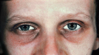

Fig. 2.29
Periorbital shadowing (“halo eyes,” “disco eyes”)
Pathophysiologically swellings and congestion of postcapillary venules in the area of the nasal sinuses have been incriminated. Occasionally a secondary hyperpigmentation on the basis of existing chronic eczema has been observed; this, however, does not correspond to a stigma. Together with the common pallor of the face, this stigma can be regarded as imbalance of the autonomic nervous system with increased alpha-adrenergic and cholinergic impulses and decreased beta-adrenergic reactions (Korting 1954).
2.3.5 Rarefaction of Lateral Eyebrows (Hertoghe)
This sign has been first described at the beginning of the twentieth century by Hertoghe in a patient with hypothyreosis. In patients with atopic eczema, some authors see an association to increased rubbing of eyebrows, when eyebrow eczema can be diagnosed at the same time (Fig. 2.30). For a long time it was regarded as a classic stigma of altered skin independent of immune reactivity. Saurat observed a regrowth of lateral eyebrow hair after successful bone marrow transplantation in patients with Wiskott-Aldrich syndrome with massive atopic eczema and atopy stigmata (Saurat 1985). It is obvious that artificial manipulations (plucking of eyebrows) (pseudo-Hertoghe) have to be differentiated from the true stigma.
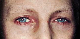

Fig. 2.30
Lateral part of eyebrows with sparse hair (“Hertoghe sign”)
2.3.6 Low Temporal Hairline, “Fur Cap Hair Growth”
This stigma describes a decreased distance between the temporal hairline and the end of the lateral eyebrows; sometimes there is no distance in between (Fig. 2.31). Quantitatively the distance between the temporal hairline and eyebrow should be more than 3 cm. The stigma can be observed in almost 90 % of patients with atopic dermatitis (Przybilla et al. 1991).
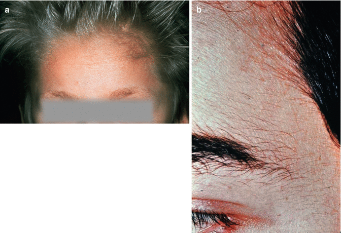

Fig. 2.31
Low temporal hairline (“fur cap”). (a) Reduction of the distance between temporal and hair and end of lateral eyebrows. (b) Low temporal hairline
A positive Hertoghe phenomenon does not exclude a low temporal hairline sign.
2.3.7 White Dermographism
The white dermographism is one of the best-known stigmata of atopy. While in healthy individuals tangential pressure (e.g., with a spatula) leads to a redness of the skin within 30–60 s (red dermographism), in atopic individuals sometimes the skin turns white (Fig. 2.32); sometimes there is a contrasting whitening next to a central red part (red dermographism with a white edge). Pathophysiologically most likely an increased local vasoconstriction is the basis of white dermographism (Wong et al. 1984).
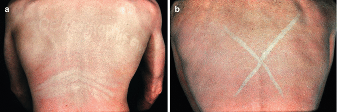

Fig. 2.32
White dermographism. (a) After tangential pressure the skin turns white instead of red. (b) Massive whitening within 30–60 s
2.3.8 Delayed Blanch
After injection of acetylcholine (intradermal), some patients with atopic eczema develop a long-lasting white discoloration in the injection area called delayed blanch reaction or delayed white reaction (Hanifin and Lobitz 1977; Lobitz and Campbell 1953; Whitfield 1938).
White dermographism and delayed blanch can be regarded as signs of autonomic dysregulation in atopics. They are not specific for atopic dermatitis but also can be seen in respiratory atopy.
It is important not to elicit white dermographism in an involved or inflamed skin area. On inflamed skin, white dermographism can be elicited also in other skin conditions.
2.3.9 Role in Diagnosis
Many studies have investigated the prevalence of atopy stigmata in patients with atopic eczema and other atopic diseases as well as in healthy control persons. In an intense and quantitative measurement in patients with various atopic diseases and healthy controls, we found that all of the abovementioned stigmata were significantly more prevalent in atopics compared to non-atopic controls (Przybilla et al. 1991). The strongest association with atopic eczema (and significantly higher than in respiratory atopy) were:
Dry skin
Ichthyosis hands
White dermographism
Hertoghe phenomenon
Similar to the increased tendency to IgE production (see Sect. 1.3.1), there also may be overlaps in the manifestation of atopy on the skin with “minimal” or “latent” atopy which only become manifest in stigma or presence of stigmata without ever turning into actual clinical eczema.
2.3.10 Summary
Stigmata have to be distinguished from actual symptoms of disease. They are signs of the skin organ not necessarily connected with feeling ill which give hints to the existence of an “atopic” diathesis. They comprise dry skin (xerosis, sebostasis), hyperlinearity of the palms and soles (ichthyosis “hands” or “feet”), increased infraorbital fold (atopy fold Dennie-Morgan), facial pallor with periorbital halo, rarefaction of lateral eyebrows (Hertoghe), fur cap hair growth with low temporal hairline, as well as white dermographism and delayed blanch.
2.4 Differential Diagnoses
Even if the diagnosis of atopic dermatitis is not difficult for an experienced dermatologist or allergist, a variety of differential diagnoses have to be considered, partly depending on the age group.
The most common differential diagnoses are listed in Table 2.2 and comprise chronic inflammatory skin diseases, infectious skin diseases, immunopathologies, hereditary skin diseases, metabolic disorders, malignant skin diseases, drug reactions, as well as all other forms of dermatitis or eczema (see also Fölster-Holst et al. 2004; Rajka 1989; Ring et al. 2006).
Table 2.2
Differential diagnoses of atopic dermatitis
Chronic inflammatory skin diseases |
Seborrheic dermatitis |
Psoriasis |
Lichen simplex chronicus |
Allergic contact dermatitis |
Irritative toxic contact dermatitis |
Infectious skin diseases |
Impetigo contagiosa |
Candidiasis |
Tinea |
Scabies |
Ictus insectorum (strophulus) |
Immunodeficiency syndromes |
Ataxia telangiectasia |
Wiskott-Aldrich syndrome |
Hyper IgE syndrome |
Severe combined immunodeficiency (SCID) |
Autoimmune diseases |
Bullous pemphigoid |
Pemphigus foliaceus |
Dermatitis herpetiformis |
Dermatomyositis |
Graft-versus-host disease |
Genodermatoses |
Netherton syndrome |
Dubowitz syndrome |
Erythrokeratodermia variabilis |
Metabolic diseases |
Phenylketonuria |
Tyrosinemia |
Zinc deficiency |
Side effects of drugs |
2.4.1 Differential Diagnosis in Infants
In the first months of life, it is mainly seborrheic dermatitis or diaper dermatitis which has to be considered (Fig. 2.33). Often there is no clear-cut diagnosis in the first months of life, and then the term “eczema infantum” (infantile eczema) is helpful (Edgren 1943).
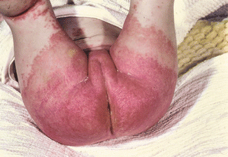

Fig. 2.33
Diaper dermatitis with marked erythema in the diaper area
There is a valuable rule: bipolar occurrence (especially head and scalp and genital area involved) allures seborrheic dermatitis (Fig. 2.34). Skin changes in the head and trunk with the uninvolved diaper area are typical for atopic eczema. This commonly observed “diaper phenomenon” is poorly investigated scientifically. One can speculate whether the diaper offers protection against scratching or the contents act like a wet wrap. It is surprising that in severely affected infants with atopic eczema, often only the diaper area is totally uninvolved (Fig. 2.35).
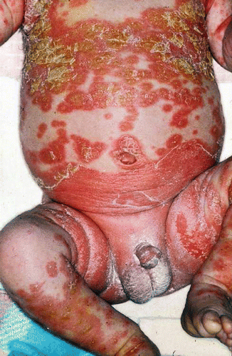


Fig. 2.34
Yellowish crusts in seborrheic dermatitis in an infant

Fig. 2.35
Diaper sign in atopic dermatitis: the diaper area is surprisingly often uninvolved. (a) Uninvolved diaper area. (b) Uninvolved diaper area with strong general skin involvement
Another sign visible to everybody but not often recognized is the absence of eczema on the tip of the nose (Yamamoto sign, personal communication via Kristian Thestrup-Pedersen 2005).
2.4.2 Chronic Inflammatory Skin Diseases
In infants, but also in small children, psoriasis can be a difficult differential diagnosis, especially in cases of so-called figurated eczema or when there is a coincidence of both diseases. Thirty years ago this was no problem; eczema and psoriasis seemed to exclude each other. Only by the epidemiological studies by Henseler and Christophers (1995), it became clear that psoriasis and atopic eczema can occur in one and the same individual, although rarely. Due to the increase in eczema prevalence, this subgroup of patients may be seen more often.
Furthermore existing eczema may trigger latent psoriasis via scratching and mechanical traumatization in the sense of a Koebner phenomenon.
A difficult differential diagnosis can be the localized involvement of palms and soles together with dyshidrosis (pompholyx). This type of hand eczema can occur in atopic eczema, but also in allergic contact dermatitis, and also in patients with tinea. According to some authors, dyshidrotic hand or foot eczema always corresponds to atopic dermatitis and should be called atopic palmoplantar eczema (Schwanitz 1992).
The co-occurrence or absence of other inflammatory diseases in atopic dermatitis is an interesting phenomenon and gives rise to new pathophysiological concepts (see Chap. 3).
Stay updated, free articles. Join our Telegram channel

Full access? Get Clinical Tree


