(1)
Department of Dermatology, Drexel University, Philadelphia, Pennsylvania, USA
Abstract
This chapter presents a pictorial representation of the variations of atopic dermatitis featured in this work, including facial-extensor, flexural, and nummular. Less frequently occurring variants are also represented, including lichen planus–like eczema, dyshidrotic eczema, pityriasis alba, and juvenile plantar dermatosis. Images also include diseases in which eczema is a known component (Meyerson’s nevus and Doucas Kapetanakis pigmented purpuric dermatosis) and diseases in which eczema was previously not thought to be present (seborrheic dermatitis, tinea pedis, and axillary granular parakeratosis).
Keywords
Atopic dermatitisAxillary granular parakeratosisDoucas Kapetanakis pigmented purpuric dermatosisDyshidrotic eczemaFacial-extensor eczemaFlexural eczemaJuvenile plantar dermatosisLichen planus–like eczemaMeyerson’s nevusNummular eczemaPityriasis albaSeborrheic dermatitisTinea pedisThis chapter presents a pictorial representation of the variations of atopic dermatitis that are featured in the body of this work. Heavily represented are the most common subtypes of atopic dermatitis: facial-extensor (Figs. 1.1, 1.2, 1.3, 1.4, and 1.5), flexural (Figs. 1.6, 1.7, 1.8, and 1.9), and nummular (Figs. 1.8, 1.9, 1.10, 1.11, 1.12, and 1.13). Less frequently occurring variants (there may not be general agreement that these are atopic dermatitis) include lichen planus–like eczema (Figs. 1.14, 1.15, and 1.16), dyshidrotic eczema, pityriasis alba (Figs. 1.17, 1.18, and 1.19), and juvenile plantar dermatosis. Diseases in which eczema is a known component, such as Meyerson’s nevus (Fig. 1.21) and Doucas Kapetanakis pigmented purpuric dermatosis (Fig. 1.20), and diseases in which eczema was previously not thought to be present, including seborrheic dermatitis (Figs. 1.22 and 1.23), tinea pedis (Figs. 1.24 and 1.25), and axillary granular parakeratosis (Fig. 1.26), are also shown.
Each of these disorders may overlap with others, so many times they are not “pure” forms of the variant. This happens relatively frequently in the facial-extensor variety presenting with flexural involvement, or with facial extensor concomitantly occurring with nummular disease. Pityriasis alba occurs very frequently with all of the major forms as well as occurring in a “solo” fashion. Dyshidrotic eczema may occasionally overlap (in my observations, most often with flexural disease). Other variants are rare enough that overlap with other forms of the disease is very uncommon. In addition to overlap, each of these may become impetiginized, with certain exceptions, such as pityriasis alba. The images are mostly from our patients at Drexel Dermatology; some have been graciously supplied by the Wake Forest Department of Dermatology.
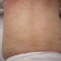
Facial
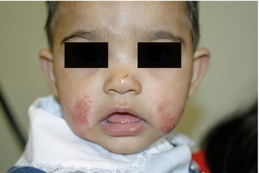
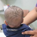
Fig. 1.1
Atopic dermatitis, facial-extensor. In this infant, there are crusted, pink-red papulovesicular plaques on bilateral cheeks
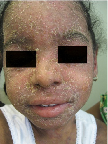
Fig. 1.2
Atopic dermatitis, facial-extensor. Marked yellowish scaling covering the entire face, and underlying pink edematous plaques, are present in this child
Fig. 1.3
 Physiology
 Immunology
 Diseases in Which Eczema Is a Secondary Component (Meyerson’s Nevus and Doucas Kapetanakis Pigmented Purpuric Dermatosis)
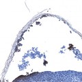
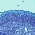
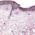 Pathology
 The Story of Eczema in Pictures
 Diseases with Occluded Sweat Ducts other than Eczema (Tinea Pedis, Axillary Granular Parakeratosis, and Seborrheic Dermatitis)


Related posts:
Physiology
Immunology
Diseases in Which Eczema Is a Secondary Component (Meyerson’s Nevus and Doucas Kapetanakis Pigmented Purpuric Dermatosis)
Pathology
The Story of Eczema in Pictures
Diseases with Occluded Sweat Ducts other than Eczema (Tinea Pedis, Axillary Granular Parakeratosis, and Seborrheic Dermatitis)
Stay updated, free articles. Join our Telegram channel
Full access? Get Clinical Tree
