Fig. 2.1
Atopic dermatitis of the antecubital fossa
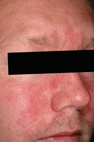
Fig. 2.2
Seborrheic dermatitis of the face
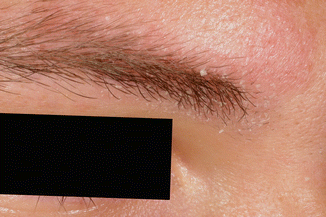
Fig. 2.3
Seborrheic dermatitis of the medial aspect of the eyebrow
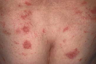
Fig. 2.4
Nummular dermatitis
By definition, contact dermatitis is the result of interaction between the skin and an object in contact with the skin. Contact dermatitis can be irritant or allergic. Protein contact dermatitis is a less common variant (see Chap. 8) as are phototoxic and photoallergic contact dermatitis (see Chap. 7). In sensitized persons, systemic presentation of the hapten may cause systemic contact dermatitis.
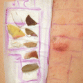
In addition to the history of the patient and a physical examination, a diagnosis of allergic contact dermatitis or protein contact dermatitis is based on the results of cutaneous testing. Patch testing is the basic test method in diagnosing allergic contact dermatitis (see Chap. 3). Test batteries are designed to take into account the most common allergens in a specific geographical region (see Chap. 5).
In addition to patch testing, prick tests and prick-prick testing may be indicated in selected cases of suspected protein contact dermatitis (see Chap. 8). In vitro test methods such as lymphocyte proliferation tests have not to date been shown to be useful routine test methods in diagnosing allergic contact dermatitis.
In most cases, it is not possible to distinguish between irritant and allergic contact dermatitis on clinical grounds. The diagnosis of irritant contact dermatitis is based on the history of the patient, the pattern of exposure, the physical findings, and the exclusion of allergic contact dermatitis.
It should be stressed that persons with seborrheic dermatitis, atopic dermatitis, or nummular eczema may develop secondary contact sensitization, thus complicating the fundamental diagnosis [1].
2.2 Clinical Features
Pruritus is the predominant symptom of contact dermatitis. Stinging with few physical signs may be experienced, and phototoxic reactions may be accompanied by a severe burning sensation.
Onset of symptoms is from seconds to minutes in contact urticaria to hours or days in contact dermatitis.
Acute contact dermatitis is characterized by erythema, edema, infiltration, and possibly vesicles (Fig. 2.5) in an area of skin that has been in contact with either an irritant or a substance to which the person has developed contact allergy. Chronic dermatitis is characterized by erythema, infiltration, scaling, and fissures (Fig. 2.6).
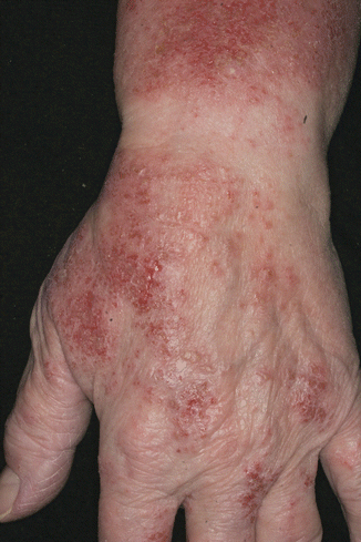
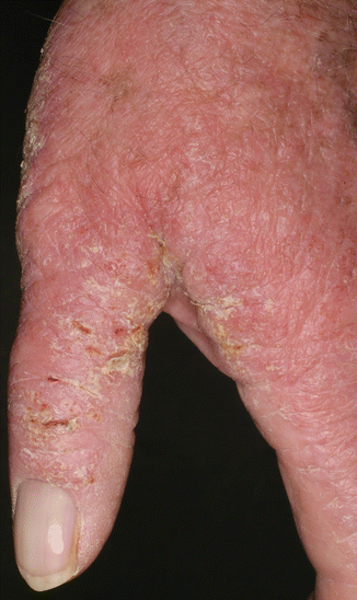
Fig. 2.5
Acute irritant contact dermatitis on the dorsal aspect of a hand
Fig. 2.6
Chronic contact dermatitis on a hand
A contact pattern is the classic clinical manifestation of contact dermatitis. The most common example of this pattern is nickel dermatitis. Nickel dermatitis develops in areas of the skin in direct contact with nickel-plated objects such as buttons, clasps, buckles (Fig. 2.7), or cell phones with nickel-plated keys (Fig. 2.8).
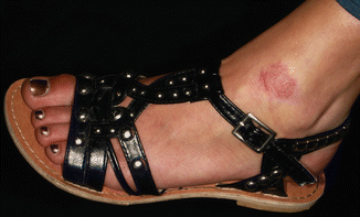
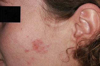
Fig. 2.7
Allergic contact dermatitis caused by nickel in the metal buckle on a sandal
Fig. 2.8
Allergic contact dermatitis caused by nickel in a mobile phone
Exposure patterns to nickel may not be as obvious as in typical allergic nickel contact dermatitis. Nickel dermatitis caused by cell phones with nickel-plated keys may, for example, be seen on the cheeks, on the sternum if the phone is carried in a pouch around the neck, or on the thigh if the phone is carried in a trouser pocket.
Airborne contact dermatitis is another clinical pattern of contact dermatitis. Dust, vapors, or fibers may cause dermatitis, typically seen on the exposed skin of the face, in particular on the eyelids, the sides and back of the neck, the forearms, and/or the dorsal aspects of the hands (Fig. 2.9) [2].
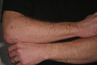
Fig. 2.9
Airborne irritant contact dermatitis caused by glass fiber used in a wind turbine wing factory
Connubial dermatitis is a term used to describe secondary exposure from a spouse or from mother to child (Fig. 2.10).
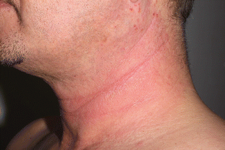
Fig. 2.10
Connubial contact dermatitis caused by contact allergy to para-toluenediamine in a hair dye used by the spouse
A striped pattern of dermatitis can be caused by irritant liquids running down an arm or a leg. A more common cause of a striped pattern of dermatitis is phototoxic contact dermatitis from plants that brush against the skin (Fig. 2.11). Phototoxic dermatitis is often vesicular or bullous and painful. The diagnosis of phototoxic contact dermatitis from plants is usually fairly obvious from the pattern of exposure (see more about plant dermatitis in Chap. 22). Unusual histories include phototoxic contact dermatitis around the mouth caused by repeated sucking on a lime and rashes in children exposed to rue rubbed on the skin to protect against mosquito bites.
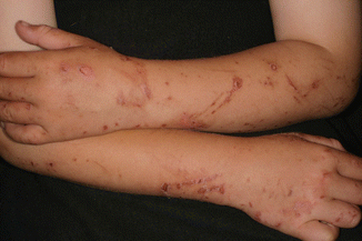
Fig. 2.11
Phototoxic dermatitis caused by contact with wild parsnip
Contact dermatitis in areas with loose subcutaneous tissue often becomes edematous. One example is edema of the eyelids following contact dermatitis of the scalp and forehead (Fig. 2.12). Another example is edema of the penis associated with contact dermatitis in the pubic area.
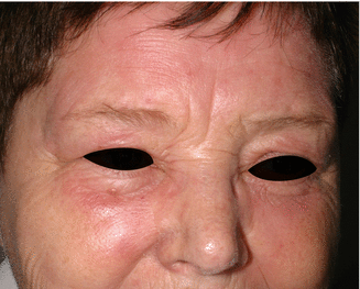
Fig. 2.12
Periorbital edema caused by allergic contact dermatitis to para-toluenediamine in hair dye
The clinical features of contact dermatitis include less common manifestations [3]. Purpuric contact dermatitis may occur following sensitization to N-isopropyl-N-phenyl-paraphenylenediamine (IPPD) used as an antioxidant in rubber or after sensitization to some textile dyes as well as to balsam of Peru. Irritant contact dermatitis from fiberglass may also induce purpura.
Textile dyes and optical whiteners have also been described as the cause of pigmented contact dermatitis [4]. It is not always possible to identify the cause of pigmented contact dermatitis (Fig. 2.13).
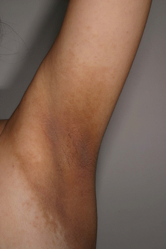
Fig. 2.13
Pigmented contact dermatitis. The specific cause was not identified in spite of extensive patch testing
Color developers have caused lichenoid contact dermatitis with lichen planus-like morphology. Hyperpigmentation follows resolution of the dermatitis. While, initially, positive patch tests have the usual morphology, they may later become lichenoid. Contact stomatitis of the oral mucosa may be lichenoid.
Erythema multiforme-like eruptions are occasionally seen, particularly in connection with allergic contact dermatitis caused by strong allergens such as certain exotic woods and other plants and para-phenylenediamine. Characteristically, intense contact dermatitis initially appears at the site of contact. After 1–2 weeks, erythema multiforme-like eruptions appear (Fig. 2.14). The eruption fades once the contact allergen is removed. A short course of systemic steroid may be useful in hastening the resolution.
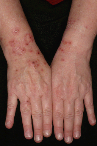
Fig. 2.14
An erythema multiforme-like reaction caused by allergic contact dermatitis
In rare instances, contact dermatitis can present as lymphomatoid tissue reactions with clinical features similar to parapsoriasis en plaques. The most common causes of this clinical manifestation are para-phenylenediamine, gold, and ethylenediamine.
2.3 Regional Contact Dermatitis
In the following, contact dermatitis typically seen in specific areas of the body (the scalp and face, the trunk, the hands, and the feet) will be described in greater detail (see Table 2.1).
Table 2.1
Common irritants/allergens
Irritant | Allergic |
|---|---|
Face and scalp | |
Hair bleaching products | Fragrances |
Fibers (rock wool, glass wool, plants) | Hair coloring agents |
Topical drugs for treatment of acne | Preservatives |
Topical drugs for treatment of actinic keratosis | |
Trunk | |
Fibers in clothing | Dyes in clothing |
Urine and feces (stoma dermatitis and diaper dermatitis) | Nickel in clasps and buckles |
Cream soaps (misuse) | Rubber additives in undergarments |
Hands | |
Detergents | Fragrances |
Wet work | Thiurams |
Soluble oils | Potassium dichromate |
Disinfectants | Preservatives |
Cement | |
Foods (vegetables, fruit, fish, meat) | |
Feet | |
Cement | Potassium dichromate |
Wood dust | Mercaptobenzothiazole |
Wet work | Dimethyl fumarate |
2.3.1 Irritant Contact Dermatitis of the Scalp
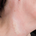
Contact dermatitis of the scalp can be either irritant or allergic. There is often more visible dermatitis at the periphery of the scalp, on, for example, the forehead and/or the neck (Fig. 2.15).
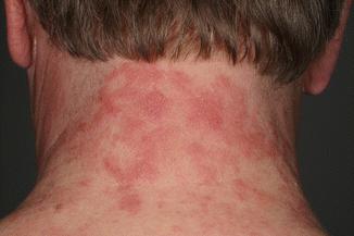
Fig. 2.15


Irritant contact dermatitis caused by a hair care product. The dermatitis was more pronounced on the neck than on the scalp itself
Related posts:


Stay updated, free articles. Join our Telegram channel
Full access? Get Clinical Tree
