Fig. 1
Onychogryphosis
Common Nail Disorders in Older Adults
In addition to the intrinsic physiologic changes of the nail plate that occur with aging, nail disorders are common in geriatric populations. This high prevalence of nail disease in the elderly is multifactorial: impaired circulation, increased susceptibility of the nail apparatus to infection due to barrier defects, increased rate of cutaneous neoplasms, elevated prevalence of comorbid dermatologic or systemic disease, and medication use are all contributing factors [2].
Brittle Nail Disease
While largely a cosmetic concern, nail brittleness is a common complaint of elderly individuals, occurring in an estimated 35 % of individuals older than 60 years [23]. Typically, affected individuals complain of soft, easily torn or split nails, and a general inability to grow longer nails [4]. As described above, longitudinal nail splitting occurs commonly in the setting of intrinsic age-related onychorrhexis (Fig. 2). Onychoschizia—or the distal lamellar peeling and splitting of the dorsal aspect of the nail plate—also is common in the elderly, particularly women, as is trachyonychia—an opaque roughness affecting the surface of the dorsal nail plate.
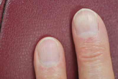
Fig. 2
Onychorrhexis
Previously, brittle nails were believed to result from decreased water content. However, this theory has since been disproved, with emerging data now demonstrating no significant difference in water content between normal and brittle nails [24]. Instead, water binding capacity has been identified as the likely causative factor, as it is reduced by nearly one half in brittle nail plates [25, 26]. As keratin, keratin-associated proteins, and lipid-content all contribute to nail plate water binding capacity, an abnormality in one or several of these factors may underlie brittle nail pathophysiology [27, 28]. Environmental factors may also further exacerbate brittle nails clinically, though they are not believed to represent a causative factor [29]. Patients with brittle nails are advised to avoid repeated hydration and desiccation of the nail plates (which often occurs occupational settings) as well as exposure to dehydrating agents used in nail cosmetics, as these may damage intracellular corneocyte bridges and dissolving intracellular lipids, thereby further increasing nail plate fragility [29, 30]. Rarely, brittle nails may also represent a harbinger of an underlying dermatologic or systemic disease, including psoriasis, lichen planus, lichen striatus, alopecia areata, Darier’s disease, peripheral arterial disease, arteriosclerosis, microangiopathy, Raynaud’s disease, polycythemia vera, dyserythropoietic anemia, thyroid disease, hypopituitarism, gout, osteoporosis, diabetes, malnutrition and nutritional disorders, chronic renal failure, hemodialysis, osteomalacia, acromegaly, pulmonary tuberculosis, chronic obstructive pulmonary disease, sarcoidosis, systemic amyloidosis, and various visceral malignancies—often in the setting of acrokeratosis paraneoplastica [31–36].
Treatment of brittle nails is often difficult given the multifactorial nature of the disease. As stated previously, environmental exposures may exacerbate any underlying nail disease, and patients should be advised to reduce frequency of prolonged contact with water, as well as detergents, cleaning solvents, and alcohol-based hand sanitizers [30]. For prolonged contact with water, cotton-lined latex gloves should be worn. Hydration of the nail plate is also essential and patients should be advised to apply a thick emollient to the entire nail plate and proximal nail fold after soaking the tips in lukewarm water for 10–20 min [37]. Urea and lactic acid have been reported as particularly effective, as both agents act to increase the water content binding capacity of the nail plate [30]. Nail cosmetics may be cautiously recommended, as once-weekly application of nail lacquer may limit exposure of water and other offending detergents, as well as reduce water vapor loss [38]. However, since acetone, and to a lesser degree acetate, dehydrate the nail plate and reduce the intrinsic lipid content, polish removers should be used on a very limited basis [38]. Cosmetic nail hardeners should also be recommended judiciously, as they contain toluene-sulfonamide-formaldehyde resins, which induce additional cross-linking between nail plate keratin. While such bonds may help to stabilize weakened nails, overuse may result in accumulation of excess keratin cross-links, thus paradoxically decreasing nail plate flexibility and increasing brittleness [38].
More recently, two new prescription medications targeting brittle nails have been released. The first agent recently gained FDA approval as a medical device for the treatment of brittle nails, and consists of a hydrosoluble nail lacquer containing hydroxypropyl chitosan, Equisetum avense extract and methylsulfonyl methane, which is marketed under the trade name Genadur ® (distributed by Medimetriks). In one study, daily applications of this agent significantly reduced longitudinal groves and lamellar splitting after 1 month [39]. The second agent, a 16 % urea polyurethane lacquer, marketed as Nu-Vail ® (distributed by Innocutis) also recently gained FDA approval as a medical device for the treatment of dystrophic nails, and was found to demonstrate a 60 % improvement in clinical parameters after 6 months of daily use [40]. Biotin supplementation can also often recommended as a systemic agent for the treatment of brittle nails, as elevated doses have been suggested to upregulate the synthesis of lipid molecules in the nail matrix, thus facilitating binding between nail plate keratinocytes [41]. Typically, dosages of 2.5–5 mg/day are recommended, as one study demonstrated a 25 % increase in nail thickness and an overall decrease in lamellar splitting with this agent [41]. Tazarotene 0.1 % cream has also anecdotally been suggested as a possible therapy, though few data exist [42].
Onychomycosis and Its Treatment in Older Adults
Onychomycosis is defined as a fungal infection that affects one or more components of the nail apparatus, with dermatophytes, yeasts, and non-dermatophytes molds all recognized as causative pathogens [43]. While the prevalence of such infections is approximately 10 % in the general population, upwards of 40–60 % of individuals over 60 years of age are affected [44, 45]. This overrepresentation of onychomycosis in geriatric populations is likely multifactorial, with reduced peripheral circulation, slower nail plate growth, inactivity, relative immunosuppression, glucose intolerance, larger distorted nail surfaces, difficulty with routine nail care and hygiene, increased risk of nail injury, and increased exposure to pathogenic fungi all representing contributing etiologic factors [3, 46].
Onychomycosis has a number of well-described common clinical presentations [47]. Of these, distolateral subungual onychomycosis is by far the most common, and is frequently associated with Trichophyton spp. (particularly T. rubrum and T. mentagrophytes). In this entity, the point of fungal entry is either the hyponychium or the lateral nail bed, resulting in the classic clinical presentation of distal onycholysis, subungual hyperkeratosis with an accumulation of subungal debris, and a thickened nail plate. Conversely, proximal subungual onychomycosis is the rarest clinical variant, and is most often associated with underlying human immunodeficiency virus infection [48]. Trichophyton rubrum is the most common causative organism associated with this condition [49]. As the infection originates at the proximal nail fold and spreads distally, clinically presenting as an opaque white discoloration of the proximal nail plate immediately overlying the lunula [43]. White superficial onychomycosis, a third clinical variant, involves the dorsal surface of the nail plate, and presents with coalescing opaque patches. The most common associated pathogen, Trichophyton mentagrophytes, contains enzymes capable of degrading nail plate keratins, so that affected areas appear crumbled and dilapidated [43].
Though many clinicians initiate treatment of onychomycosis based on clinical impression alone, this is not recommended as many other entities present with similar clinical findings. Pathologic diagnosis can easily be obtained through periodic-acid Schiff (PAS) staining of nail clippings or subungual debris, though it cannot reliably speciate the infecting organisms. Additionally, the pathologic diagnosis relies on the presence of branching septae; yeast forms alone do not confirm fungal infection of the nail apparatus [50]. Culture remains the gold standard for diagnosis, as it also identifies the pathogenic organism and allows antifungal sensitivity testing, though has a relatively low sensitivity of 53 % [51]. Accordingly, best practice guidelines dictate that both culture and PAS staining should be performed, as this combination yields a sensitivity of 96 % [51].
Treatment of onychomycosis in elderly populations has traditionally been difficult, primarily due to poor response, frequent relapses, and elevated associated risk profiles [3]. Topical therapy definitively carries the lowest risk of toxicity, though has historically not been curative. Instead, topical treatments have often been employed as a palliative measures, preventing the spread of fungal infection to neighboring nails. Ciclopirox 8 % lacquer is the most well studied topical agent, demonstrating clinical cure in 5.5–8.5 % of all cases [52]. Urea 40 % gel has also been anecdotally recommended as an adjunctive agent, theoretically increasing cutaneous absorption of any topical antifungal preparation [53]. However, the recent FDA approval of two new topical agents for will likely affect the treatment algorithm for onychomycosis, particularly in elderly adults.
Efinaconazole (Jublia ® distributed by Valeant) is a new triazole antifungal topical solution that received FDA approval in June 2014 for the treatment of onychomycosis. Data from duplicate industry-sponsored phase III clinical studies demonstrated that 17.8 and 15.2 % of all study subjects attained a complete cure after 52 weeks of daily treatment [54]. An additional 35.7 and 31.0 % of study subjects were also noted to have less than 10 % of nail plate involvement after completing the treatment regimen. The most common attributable adverse reactions reported during the preclinical studies were application site dermatitis (3.5 %), application site vesicles (2.0 %, 1.2 %), contact dermatitis (2.9 %, 1.4 %) and ingrowing nail (2.6 %, 1.9 %), though one subsequent smaller study suggests the ability of efinaconazole to induce delayed contact sensitivity may be limited [54, 55].
Tavaborole (Kerydin ® distributed by Anacor Pharmaceuticals), a first-in-class, boron-containing oxaborole broad-spectrum with antifungal activity against dermatophytes, yeast, and non-dermatophyte molds, also recently received FDA approval for the treatment of onychomycosis [56]. In duplicate industry-sponsored phase III clinical trials, complete clinical cure was achieved 6.5 and 9.1 % of patients treated with tavaborole for 360 days [57]. An “almost clear nail” was also observed in an additional 26.1 and 27.5 % of study subjects. In both the phase II and phase III clinical trials, adverse events were infrequent and limited to cutaneous irritation [57].
Regardless of the topical agent selected, the infected portion of the nail plate should be frequently debrided, ideally every 3–4 weeks [4]. Removal of the affected nail plate and subungual debris not only decreases fungal load, but also may increase topical therapy penetration [58]. While topical therapies carry a very low risk of toxicity, compliance with daily or twice daily applications can be difficulty for elderly individuals, particularly in the setting of limited mobility or other musculoskeletal or rheumatologic comorbidities [59].
Surgical or chemical nail avulsion represents an additional adjunctive therapy that best utilized in cases with lateral nail plate involvement [60]. The presence of a dermatophytoma—a subungual mass of densely packed fungal hyphae with poor antifungal penetration—is another indication for partial or complete nail debridement [60, 61].
Systemic antifungal therapy remains the gold standard for treatment of onychomycosis. Terbinafine, fluconazole, and itraconazole are the three agents most widely prescribed systemic antifungals. Due to the safety and efficacy concerns, older agents such as griseofulvin and ketoconazole are infrequently prescribed. Of the former three, only terbinafine and itraconazole are FDA-approved for the treatment of onychomycosis. Prior to the initiation of any systemic treatment regimen, the following factors should be considered: the patient’s relevant past medical history, current medications, and the causative or suspected pathogen.
Terbinafine is a synthetic allylamine antifungal with fungicidal activity against fungi, dermatophytes, and some yeast forms through the inhibition of squalene epoxidase [62]. This agent is currently the drug of choice for treating onychomycosis, as it has been repeatedly demonstrated to have the highest rates of clinical cure and the lowest rates of recurrence [63–65]. Terbinafine is also highly lipophilic and persists in the nail plate for several months after discontinuation [62]. Terbinafine is dosed at 250 mg daily for 6 weeks for fingernail onychomycosis and 12 weeks for toenail onychomycosis. The data regarding the overall efficacy rate for terbinafine vary. Several meta-analyses suggest a clinical cure rate between 66 and 75 %, though early studies demonstrate rates of 38–54 % [65–69]. Adverse effects commonly attributed to terbinafine include nausea, gastrointestinal disturbance, dysgeusia, leukopenia, liver function abnormalities, and cutaneous eruption [43]. Rarely, terbinafine has been associated with fulminant liver failure, and thus, intermittent laboratory assessment of hepatic parameters is often performed. Terbinafine has few significant drug interactions, and can be co-administered statins, digoxin, warfarin, and many other medications commonly prescribed in elderly populations [43]. However, this agent is a potent inhibitor of CYP2D6, affecting the metabolism of numerous medications including many beta-blockers as well as donepezil, and thus, medication reconciliation and review is recommended prior to initiating therapy [70]. In older patients who may be on multiple medications (including hepatically metabolized drugs such as statins), pulse dose treatment consisting of 7 days a month for 4 months has been shown to reduce likelihood of transaminitis with only slightly lower cure rates [67].
Itraconazole is a synthetic triazole antifungal, and has a broad spectrum of fungistatic activity but potential side effects with older adults should be carefully considered [71]. Through the inhibition of ergosterol synthesis, itraconazole is able to inhibit growth of dermatophytes, Candida, and some nondermatophyte molds [71]. Similar to terbinafine, itraconazole is also highly lipophilic and persists in the nail plate for 6–9 months after discontinuation [71]. Two dosing regimens exist for itraconazole: daily treatment and intermittent (pulse) therapy. Daily treatment is dosed at 200 mg/day for 8 weeks for fingernail onychomycosis and 12 weeks for toenail onychomycosis. Pulse therapy, which is not FDA-approved for the treatment of toenail onychomycosis, consists of 400 mg/day for the first week of every month and lasts 2 months for fingernail onychomycosis and 3 months for toenail onychomycosis. As with terbinafine, the data regarding the efficacy of itraconazole vary, though most larger and head-to-head studies suggest terbinafine has a higher overall clinical cure rate than either of the itraconazole dosing regimens [65, 66, 68]. Notably, itraconazole is absolutely contraindicated in patients with a past medical history of congestive heart failure due to its negative inotropic effect [72]. Other associated adverse reactions include nausea, gastrointestinal disturbance, telogen effluvium, hepatotoxicity, and cutaneous eruption [43]. Itraconazole is also a potent inhibitor of CYP3A4, and thus interacts with many medications. Those specifically mentioned in the associated FDA-mandated black box warning include: cisapride, midazolam, nisoldipine, felodipine, pimozide, quinidine, dofetilide, triazolam, levacetylmethadol, lovastatin, simvastatin, ergot alkaloids, and methadone [73]. Given these associations and interactions, itraconazole is not typically a first-line agent in onychomycosis among elderly populations, though it has a definite clinical value in certain settings [74].
Of note, two published, peer-reviewed studies exist that specifically pertain to the treatment of onychomycosis in the elderly. The first—a single blind, randomized, non-industry-sponsored, prospective study—compared the efficacy of terbinafine and itraconazole in the treatment of onychomycosis [75]. In total, 101 individuals aged 60 years and older with dermatophytosis involving at least one hallux were enrolled. Half were treated with terbinafine 250 mg daily for 12 weeks, while the remaining individuals were treated with itraconazole 200 mg twice daily for 1 week per month for 3 months [75]. The resulting data demonstrated no significant difference in cure rates: the clinical cure rates were 62 % for terbinafine and 60.8 % for itraconazole [75]. Adverse events were noted in five individuals in the terbinafine treatment group and seven individuals in the itraconazole treatment group, all of which were mild and reversible [75]. The second study performed a subanalysis of patients 65 years and older from an open-label, randomized, multicenter study of adults treated with terbinafine or terbinafine and surgical nail debridement for onychomycosis [76]. Specifically, surgical nail debridement in the context of onychomycosis traditionally refers to the removal of all onycholytic portions of the nail plate, thus reducing the fungal load by eliminating the subungual debris [76]. The resulting data demonstrated that patients treated with both terbinafine and surgical debridement had higher rates of complete cure than those treated with terbinafine alone [76]. Additionally, the most frequently reported adverse events included nausea, arthralgia, and hypercholesterolemia, with three participants ultimately withdrawing due to a medication-related adverse effect [76]. Of the enrolled individuals 97 % of patients were concomitantly taking another oral medication throughout the course of the study: 64 % antihypertensives, 25 % antidiabetics, and 47 % lipid-lowering medications [76].
Lastly, various lasers and other light-based devices have been posited as a potential alternative therapeutic modality for onychomycosis, although their exact mechanism of action is unclear. Currently, the five devices are currently approved by the FDA for the treatment of onychomycosis, all of which are short-pulse neodymium-doped yttrium aluminum garnet (Nd:Yag) lasers [77]. Notably, all five devices only received approval for temporary increase in clear nail in patients with onychomycosis, not definitive treatment [78]. While the idea of laser treatment for onycomycosis seems promising, to date the data supporting the actual efficacy of this modality is mixed. One review of all relevant published studies pertaining to treatment of onychomycosis with the Nd:Yag reemphasized that while results were favorable, all studies were small and many poorly designed [77]. Furthermore, a more recent larger, randomized controlled trial found no significant difference in mycological culture or nail plate clearance [78].
Nail-Associated Neoplasms in the Elderly
Tumors of the nail unit can be classified as benign, malignant, or metastatic. While fibrous tumors are the most common neoplasms of the nail apparatus in the general population, digital myxoid cysts predominate as the most prevalent among the elderly [3, 79, 80]. These tumors have many synonyms, including pseudomyxoid cysts, ganglion cysts, and digital mucous cysts, focal mucinosis, periarticular fibromas, and cutaneous myxoid cysts, among others [81, 82]. Digital myxoid cysts classically present as skin-colored to blue, smooth, translucent, dome-shaped papule located distal to the distal interphalangeal (DIP) joint, most commonly on the first three fingers [3, 81]. Histopathologically, these lesions are characterized by a deep focal mucinosis on acral skin without an epithelial lining. In over 80 % of cases digital myxoid cysts connect with the underlying DIP joint [83]. In cases where the lesion is located more distally and involves the proximal nail fold, the lesion may compress the underlying nail matrix and result in a longitudinal grove in the nail plate [81]. Generally, asymptomatic digital myxoid cysts are best observed clinically. For symptomatic or cosmetically bothersome lesions, surgical excision with ligation of communicating tract to the underlying joint is the gold standard for treatment [84]. Simple drainage of the lesions is a less invasive alternative for patients unwilling or unable to undergo surgery, though it is also associated with higher rates of recurrence [85].
Glomus tumors represent another common benign neoplasm involving the nail unit. While glomus tumors can develop at any site on the body, upwards of 75 % occur on the hand, most commonly on the fingertips [81]. Epidemiologically, over 90 % of cases occur in women, with an average age of 45 years [86]. Glomus tumors have two distinct clinical presentations: (1) a small red or blue macule on the nail bed that is visible through the nail plate (Fig. 3), or (2) longitudinal erythronychia—or a longitudinal erythematous streak that extends the length of the nail bed visible through the nail plate—with an overlying furrow in the nail plate and distal nicking [87]. The characteristic triad of associated pain, pinpoint tenderness and temperature (especially cold) sensitivity is highly specific in reaching a diagnosis [87]. Magnetic resonance imaging can be used to confirm the diagnosis in most cases. This imaging modality has the best capacity to assess the size and extent of the lesion and provides the highest sensitivity amongst all imaging modalities [88–90]. Surgical excision the primary treatment for these tumors, though some evidence suggests a recurrence rate of approximately 17 % [91]. Asymptomatic lesions can be monitored clinically, though further evaluation and biopsy are recommended if squamous cell carcinoma or amelanotic melanoma is in the differential diagnosis.
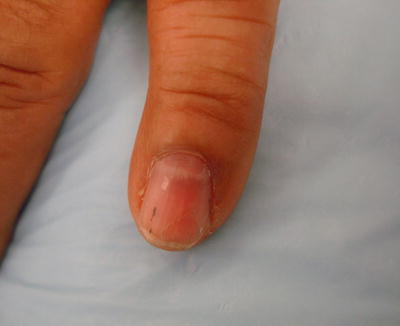
Fig. 3
Glomus tumor
Squamous cell carcinoma is the most common malignant neoplasm affecting the nail apparatus [81, 87]. While epidemiologic data for this malignancy is limited, one retrospective study found a male predominance, with a peak incidence between 50 and 69 years of age [92]. Risk factors for subungual or periungual squamous cell carcinoma include: trauma, ionizing radiation, arsenic exposure, dyskeratosis congenita, and human papillomavirus (HPV) infection [92–94]. Specifically, HPV subtype 16 DNA has been identified by polymerase chain reaction (PCR) in 74 % of all reported cases [93]. The morphologic presentation of squamous cell carcinoma of the nail unit can be variable and may mimic a variety of benign conditions clinically, often delaying diagnosis and appropriate treatment [87, 95]. While subungual involvement is most common, tumors may also arise in the paronychial epithelium [87]. Subungual lesions typically present with onycholysis overlying a verrucous or hyperkeratotic mass or a frank ulcer (Fig. 4) [87


Related posts:


Stay updated, free articles. Join our Telegram channel
Full access? Get Clinical Tree
